. 1999 Aug;9(8):751-62.

High-resolution landmark framework for the sequence-ready mapping of Xq23-q26.1

Affiliations

High-resolution landmark framework for the sequence-ready mapping of Xq23-q26.1

H E Steingruber et al. Genome Res. 1999 Aug.

Abstract

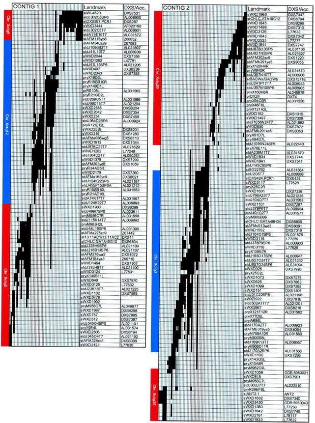
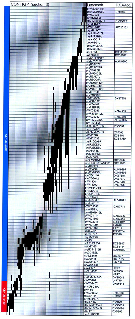
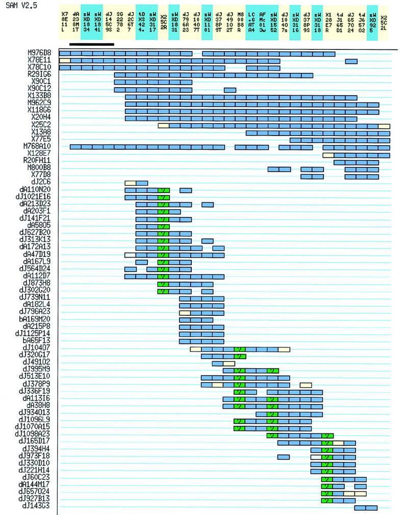
We have established a landmark framework map over 20-25 Mb of the long arm of the human X chromosome using yeast artificial chromosome (YAC) clones. The map has approximately one landmark per 45 kb of DNA and stretches from DXS7531 in proximal Xq23 to DXS895 in proximal Xq26, connecting to published framework maps on its proximal and distal sides. There are three gaps in the framework map resulting from the failure to obtain clone coverage from the YAC resources available. Estimates of the maximum sizes of these gaps have been obtained. The four YAC contigs have been positioned and oriented using somatic-cell hybrids and fluorescence in situ hybridization, and the largest is estimated to cover approximately 15 Mb of DNA. The framework map is being used to assemble a sequence-ready map in large-insert bacterial clones, as part of an international effort to complete the sequence of the X chromosome. PAC and BAC contigs currently cover 18 Mb of the region, and from these, 12 Mb of finished sequence is available.


Figures

Figure 1
Figure 1
Hybrid interval map and framework map summary. A G-banded representation of Xq22.3–q26.3. The deletion-hybrid breakpoints define nine intervals (A–I) into which the 69 STSs have been placed. Within deletion intervals, STSs and their associated loci are shown in the order in which they appear on the framework map. The approximate positions and extents of the four YAC contigs are indicated by numbered bars. Numbers within the arcs at left of the contigs indicate the number of separate FISH experiments, using different PAC probe combinations, that support the contig order (arc drawn between bars) or orientation (within bars).
Figure 2
Figure 2
The landmark framework map of Xq23–q26.1. The landmark order on the four YAC contigs is illustrated. YAC clones are shown as thin vertical bars with positive landmark content indicated in black. Cases in which YAC clones have not been tested with specific markers are indicated by checkered boxes. Contig 4 is shown in three sections. For ease of alignment, some landmarks (solid gray boxes) and YAC clones have been shown duplicated at the end and beginning of appropriate sections. Landmark STSs (st or sWXD prefix) and probes (pr) are shown in the first column, and DXS numbers, database accession numbers, or Genome Database (GDB) identifiers in the second. Published STSs are derived from the Généthon (stAFM), Whitehead Institute (stWI), Co-operative Human Linkage Center (stCHLC), Center for Genetics in Medicine (sWXD) maps and databases, from GDB (various), or from the literature (see Methods). PAC end STSs are named after the PAC clone of origin with the suffix SP6 or T7 (e.g., stdJ364D2SP6). PAC clones are derived from the Roswell Park Cancer Institute libraries RPCI1, RPCI3, RPCI4, RPCI5 (dJ), and RPCI6 (dA). YAC end hybridization probes are named after the clones from which they are derived with the “L” or “R” suffix (see Methods). Clones are from the CEPH (yM), ICI (yR), ICRF [yX (ICRFy900), yY (ICRFy901), yZ (ICRFy905)], and Washington University libraries (yA, yB), or were provided by colleagues (see Methods). Where YAC end probes have been sequenced and used to design STSs, EMBL accession numbers are shown. Three of these STSs were used for YAC library screening in preference to the end hybridization probe [stSG11439 (pryR17HB2R), stSG14848 (pryM949F11R), and stSG22782 (pryM963A8R)]. At left of each YAC contig are shown the extents of the corresponding bacterial clone contigs as red or blue boxes. Bacterial clone contigs are numbered and have the prefix Chr_Xctg (where space allows). In contig 4 (section 1), contigs X_CTG213, 377, and 104 are labeled in boxes to the right of those describing their extents because of space constraints. All of the bacterial clone contigs can be viewed in detail by following instructions at http://www.sanger.ac.uk/HGP/ChrX/. To avoid confusion, the lower numbered contig will be maintained on the WWW site as contigs join. A magnified version of this map showing YAC clone names can be viewed via http://www.sanger.ac.uk/HGP/ChrX.
Figure 2
Figure 2
The landmark framework map of Xq23–q26.1. The landmark order on the four YAC contigs is illustrated. YAC clones are shown as thin vertical bars with positive landmark content indicated in black. Cases in which YAC clones have not been tested with specific markers are indicated by checkered boxes. Contig 4 is shown in three sections. For ease of alignment, some landmarks (solid gray boxes) and YAC clones have been shown duplicated at the end and beginning of appropriate sections. Landmark STSs (st or sWXD prefix) and probes (pr) are shown in the first column, and DXS numbers, database accession numbers, or Genome Database (GDB) identifiers in the second. Published STSs are derived from the Généthon (stAFM), Whitehead Institute (stWI), Co-operative Human Linkage Center (stCHLC), Center for Genetics in Medicine (sWXD) maps and databases, from GDB (various), or from the literature (see Methods). PAC end STSs are named after the PAC clone of origin with the suffix SP6 or T7 (e.g., stdJ364D2SP6). PAC clones are derived from the Roswell Park Cancer Institute libraries RPCI1, RPCI3, RPCI4, RPCI5 (dJ), and RPCI6 (dA). YAC end hybridization probes are named after the clones from which they are derived with the “L” or “R” suffix (see Methods). Clones are from the CEPH (yM), ICI (yR), ICRF [yX (ICRFy900), yY (ICRFy901), yZ (ICRFy905)], and Washington University libraries (yA, yB), or were provided by colleagues (see Methods). Where YAC end probes have been sequenced and used to design STSs, EMBL accession numbers are shown. Three of these STSs were used for YAC library screening in preference to the end hybridization probe [stSG11439 (pryR17HB2R), stSG14848 (pryM949F11R), and stSG22782 (pryM963A8R)]. At left of each YAC contig are shown the extents of the corresponding bacterial clone contigs as red or blue boxes. Bacterial clone contigs are numbered and have the prefix Chr_Xctg (where space allows). In contig 4 (section 1), contigs X_CTG213, 377, and 104 are labeled in boxes to the right of those describing their extents because of space constraints. All of the bacterial clone contigs can be viewed in detail by following instructions at http://www.sanger.ac.uk/HGP/ChrX/. To avoid confusion, the lower numbered contig will be maintained on the WWW site as contigs join. A magnified version of this map showing YAC clone names can be viewed via http://www.sanger.ac.uk/HGP/ChrX.
Figure 3
Figure 3
Comparison of the framework map with other published maps. A comparison is shown between the landmark order on the framework map (B), the Généthon genetic map (A) (Dib et al. 1996), and the Center for Genetics in Medicine YAC–STS map (C) (Nagaraja et al. 1998). All STSs shared by the framework map and these other maps are shown. Genetic distances are shown for A, except in cases where distance is <1 cM. Boxes around STSs in B and C indicate that markers are from the same YAC contig. In the comparison of B and C, lines are drawn only when landmarks show differences in order between the two maps.
Figure 4
Figure 4
Segments of YAC contig 2 and of the underlying bacterial clone contig (Chr_Xctg3). The image is taken from the graphical display of the contig assembly program SAM. Landmarks are shown at top, with clone ends (YAC or PAC) in yellow and other STSs in blue–green. YACs are named according to Fig. 2; PACs are prefixed dJ (RPCI1, RPCI3, RPCI4, and RPCI5) or dA (RPCI6); and BACs, bA (RPCI11). Dark blue boxes indicate strong positive landmark–clone associations; and pale gray, weak associations; a yellow box indicates the end of the YAC or PAC clone from which a probe or STS was derived; green boxes show cases in which bacterial clones have not been screened for the relevant markers.

References

    1. Anand R, Riley JH, Butler R, Smith JC, Markham AF. A 3.5 genome equivalent multi access YAC library: Construction, characterisation, screening and storage. Nucleic Acids Res. 1990;18:1951–1956. - PMC - PubMed
    1. Bentley DR, Todd C, Collins J, Holland J, Dunham I, Hassock S, Bankier A, Gianelli F. The development and application of automated gridding for efficient screening of yeast and bacterial ordered libraries. Genomics. 1992;12:534–541. - PubMed
    1. Bentley DR, Pruitt KD, Deloukas P, Schuler GD, Ostell J. Coordination of human genome sequencing via a consensus framework map. Trends Genet. 1998;14:381–384. - PubMed
    1. Brownstein BH, Silverman GA, Little RD, Burke DT, Korsmeyer SJ, Schlessinger D, Olson MV. Isolation of single-copy human genes from a library of yeast artificial chromosome clones. Science. 1989;244:1348–1351. - PubMed
    1. Buetow KH, Weber JL, Ludwigsen S, Scherpbier-Heddema T, Duyk GM, Sheffield VC, Wang Z, Murray JC. Integrated human genome-wide maps constructed using the CEPH reference panel. Nat Genet. 1994;6:391–393. - PubMed

Publication types

Substances

LinkOut - more resources