

Increased inducible apoptosis in CD4+ T lymphocytes during polymicrobial sepsis is mediated by Fas ligand and not endotoxin
- PMID: 10447713
- PMCID: PMC2326799
- DOI: 10.1046/j.1365-2567.1999.00765.x
Increased inducible apoptosis in CD4+ T lymphocytes during polymicrobial sepsis is mediated by Fas ligand and not endotoxin
Abstract
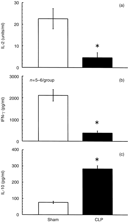
Recent studies suggest that increased lymphocyte apoptosis (Ao) detected in peripheral blood T cells from burn patients appears to contribute to decreased lymphocyte immunoresponsiveness. However, while it is known that sepsis induces a marked depression in the splenocyte immune response (i.e. decreased interleukin-2, interferon-gamma production and proliferation) in response to the T-cell mitogen concanavalin A (Con A), it is unknown whether this depression is associated with an increase in inducible Ao and if so, which mediators control this process. To assess this, splenocytes were harvested from mice at 24 hr (a period associated with decreased Con A response) after the onset of polymicrobial sepsis [caecal ligation and puncture (CLP)] or sham-CLP (Sham) and then stimulated with 2.5 microg Con A/ml (24 hr). Septic mouse splenocytes stimulated with Con A, while not showing a change in their phenotypic make-up, did exhibit a marked increase in the percentage of splenocyte that were Ao+ which was associated with altered cytokine release. This appears to be due to an increase in the percentage of Ao+ cells in the CD4+ CD8- population and was associated with enhanced Fas antigen expression as well as an increase in mRNA for the Fas-FasL gene family. To determine if the changes in Ao are due to either endotoxin (a product of Gram-negative bacteria seen in CLP mice) or the expression of Fas ligand (FasL; a mediator of activation-induced lymphocyte Ao), a second set of studies examining Con A-inducible Ao was performed with splenocytes harvested from septic endotoxin-tolerant C3H/HeJ and the FasL-deficient C3H/HeJ-Fasl gld mice. The results show that increased splenocyte Ao detected following CLP is due to a FasL-mediated process and not to endotoxin. Thus the inadvertent up-regulation of FasL-mediated splenocyte Ao may contribute to the depression of splenocyte immune responses seen during polymicrobial sepsis.
Figures


Similar articles
-
Is Fas ligand or endotoxin responsible for mucosal lymphocyte apoptosis in sepsis?Arch Surg. 1998 Nov;133(11):1213-20. doi: 10.1001/archsurg.133.11.1213. Arch Surg. 1998. PMID: 9820353
-
Does Fas ligand or endotoxin contribute to thymic apoptosis during polymicrobial sepsis?Shock. 1999 Mar;11(3):211-7. doi: 10.1097/00024382-199903000-00010. Shock. 1999. PMID: 10188775
-
Increased mucosal B-lymphocyte apoptosis during polymicrobial sepsis is a Fas ligand but not an endotoxin-mediated process.Blood. 1998 Feb 15;91(4):1362-72. Blood. 1998. PMID: 9454767
-
Antigen-induced T cell death is regulated by CD4 expression.Int Rev Immunol. 2001 Oct;20(5):535-46. doi: 10.3109/08830180109045577. Int Rev Immunol. 2001. PMID: 11890611 Review.
-
Involvement of Fas-Fas ligand interactions in graft rejection.Int Rev Immunol. 1999;18(5-6):527-46. doi: 10.3109/08830189909088497. Int Rev Immunol. 1999. PMID: 10672500 Review.
Cited by
-
A new mass spectrometry-based method for the quantification of histones in plasma from septic shock patients.Sci Rep. 2017 Sep 6;7(1):10643. doi: 10.1038/s41598-017-10830-z. Sci Rep. 2017. PMID: 28878320 Free PMC article.
-
Regulatory roles of CD1d-restricted NKT cells in the induction of toxic shock-like syndrome in an animal model of fatal ehrlichiosis.Infect Immun. 2008 Apr;76(4):1434-44. doi: 10.1128/IAI.01242-07. Epub 2008 Jan 22. Infect Immun. 2008. PMID: 18212072 Free PMC article.
-
Second hit post burn increased proximal gut mucosa epithelial cells damage.Shock. 2008 Aug;30(2):184-8. doi: 10.1097/SHK.0b013e318162a3f6. Shock. 2008. PMID: 18197149 Free PMC article.
-
Role of Mitochondrial DNA in Septic AKI via Toll-Like Receptor 9.J Am Soc Nephrol. 2016 Jul;27(7):2009-20. doi: 10.1681/ASN.2015040376. Epub 2015 Nov 16. J Am Soc Nephrol. 2016. PMID: 26574043 Free PMC article.
-
Dysregulated cytokine expression by CD4+ T cells from post-septic mice modulates both Th1 and Th2-mediated granulomatous lung inflammation.PLoS One. 2011;6(5):e20385. doi: 10.1371/journal.pone.0020385. Epub 2011 May 31. PLoS One. 2011. PMID: 21655295 Free PMC article.
References
-
- Schwartz LM, Osborne BA. Programmed cell death, apoptosis and killer genes. Immunol Today. 1993;14:582. - PubMed
-
- Cohen JJ, Duke RC, Fadok VA, Sellins KS. Apoptosis and programmed cell death in immunity. Annu Rev Immunol. 1992;10:267. - PubMed
-
- Ayala A, Herdon CD, Lehman DL, Demaso CM, Ayala CA, Chaudry IH. The induction of accelerated thymic programmed cell death during polymicrobial sepsis: Control by corticosteroids but not tumor necrosis factor. Shock. 1995;3:259. - PubMed
-
- Ayala A, Herdon CD, Lehman DL, Ayala CA, Chaudry IH. Differential induction of apoptosis in lymphoid tissues during sepsis: variation in onset, frequency, and the nature of the mediators. Blood. 1996;87:4261. - PubMed
-
- Ayala A, Deol ZK, Lehman DL, Herdon CD, Chaudry IH. Polymicrobial sepsis but not low dose endotoxin infusion causes decreased splenocyte IL-2/IFN-gamma release while increasing IL-4/IL-10 production. J Surg Res. 1994;56:579. - PubMed
Publication types
MeSH terms
Substances
Grants and funding
LinkOut - more resources
Full Text Sources
Medical
Research Materials
Miscellaneous
