

Infection-stimulated infraosseus inflammation and bone destruction is increased in P-/E-selectin knockout mice
- PMID: 10447722
- PMCID: PMC2326818
- DOI: 10.1046/j.1365-2567.1999.00754.x
Infection-stimulated infraosseus inflammation and bone destruction is increased in P-/E-selectin knockout mice
Abstract
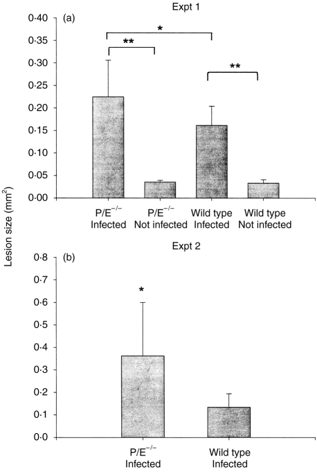
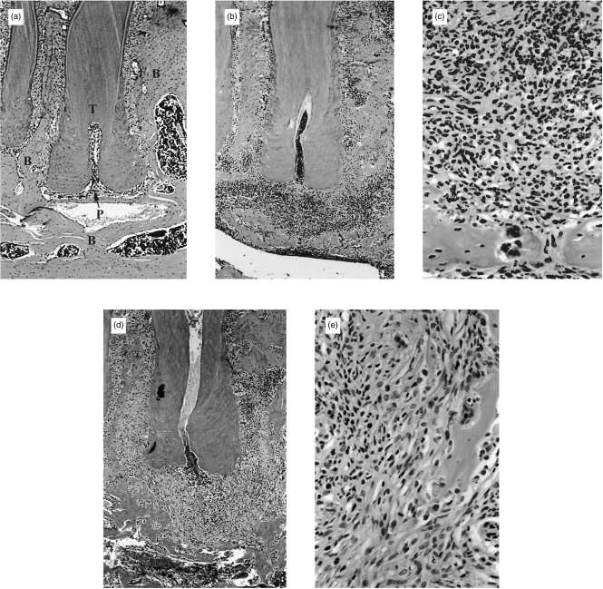
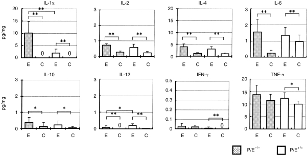
Infections of the dental pulp commonly result in infraosseus inflammation and bone destruction. However, the role of phagocytic leucocytes in the pathogenesis of pulpal infections has been uncertain. In this work we used P/E-/- selectin-deficient mice, which lack rolling adhesion of leucocytes to endothelium and mimic the human syndrome, leucocyte adhesion deficiency II (LAD-II), to test the hypothesis that phagocytic leucocytes protect against pulpal infection and subsequent periapical infraosseus bone resorption. P/E-/- mice and P/E+/+ wild-type controls were subjected to surgical pulp exposure, and both groups were infected with a mixture of pulpal pathogens including Prevotella intermedia, Fusobacterium nucleatum, Peptostreptococcus micros and Streptococcus intermedius. Animals were killed after 20 days, and the extent of infraosseus bone destruction was quantified by histomorphometry. In two separate experiments, P/E-/- mice had significantly greater bone resorption than P/E+/+ controls. The increased bone destruction correlated with a twofold decrease in polymorphonuclear (PMN) infiltration into periapical inflammatory tissues of P/E-/- mice. P/E-/- mice had higher tissue levels of the bone resorptive cytokine, interleukin (IL)-1alpha. Tissue levels of IL-2, IL-4, IL-10, tumour necrosis factor-alpha (TNF-alpha) and interferon-gamma (IFN-gamma) were all higher in P/E-/- mice, but the increases were not statistically significant. Only IL-12 was higher in P/E+/+ mice, possibly reflecting a greater number of infiltrating monocytes in wild-type mice. These findings demonstrate that phagocytic leucocytes are protective in this model, and suggest that elevated expression of inflammatory cytokines is responsible for the observed bone destruction.
Figures

References
-
- Bergenholtz G. Pathogenic mechanisms in pulpal disease. J Endon. 1990;16:98. - PubMed
-
- Bergenholtz G, Lindhe J. Effect of soluble plaque factors on inflammatory reactions in the dental pulp. Scand J Dental Res. 1975;83:153. - PubMed
-
- Okiji T, Kawashima N, Kosaka T, et al. Distribution of Ia antigen-expressing nonlymphoid cells in various stages of induced periapical lesions in rat molars. J Endod. 1994;20:27. - PubMed
-
- Kawashima N, Okiji T, Kosaka T, Suda H. Kinetics of macrophages and lymphoid cells during the development of experimentally induced periapical lesions in rat molars: a quantitative immunohistochemical study. J Endod. 1996;22:311. - PubMed
-
- Stern MH, Dreizen S, Mackler BF, et al. Quantitative analysis of cellular composition of human periapical granulomas. J Endod. 1981;7:117. - PubMed
Publication types
MeSH terms
Substances
Grants and funding
LinkOut - more resources
Full Text Sources
Medical
Molecular Biology Databases
