

Activation, survival and apoptosis of CD45RO+ and CD45RO- T cells of human immunodeficiency virus-infected individuals: effects of interleukin-15 and comparison with interleukin-2
- PMID: 10447730
- PMCID: PMC2326826
- DOI: 10.1046/j.1365-2567.1999.00807.x
Activation, survival and apoptosis of CD45RO+ and CD45RO- T cells of human immunodeficiency virus-infected individuals: effects of interleukin-15 and comparison with interleukin-2
Abstract
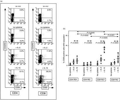
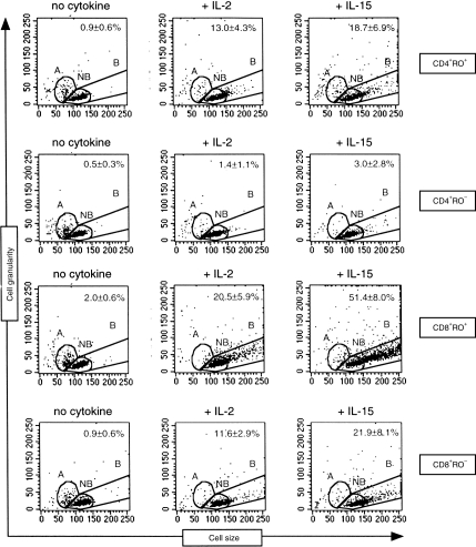
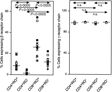
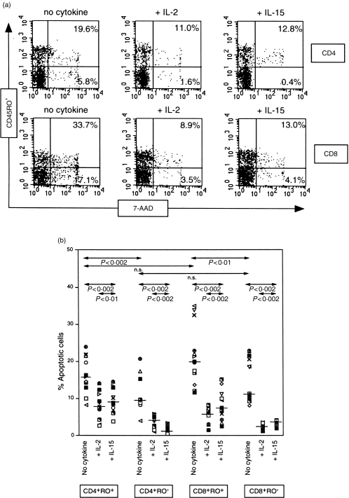
HIV infection is associated with increased representation of T cells bearing an activated, memory (CD45RO+) phenotype. Although administration of antiretroviral agents and interleukin-2 (IL-2) augment depleted CD4+ T-cell numbers, such therapies have been preferentially beneficial for CD45RO+ T cells. Interleukin-15 (IL-15) exhibits many biological activities in common with IL-2, including promoting T-cell survival and proliferation. The present study found that these two cytokines differed in their ability to induce proliferation, enhance survival, and control apoptosis of CD45RO+ and CD45RO- T-cell populations of human immunodeficiency- (HIV) infected individuals. When used at equivalent concentrations in vitro, IL-15 was more potent than IL-2 in activating and stimulating proliferation of CD4+CD45RO+, CD8+CD45RO+ and CD8+CD45RO- cells, but failed to be more effective than IL-2 in reducing apoptosis. Poor activation of CD4+CD45RO- cells by IL-15 and to IL-2 appeared to be attributable to low expression of the beta receptor chain utilized by both cytokines. However, IL-15 was more effective than IL-2 in enhancing survival of the CD4+CD45RO- population, suggesting a greater protective effect of IL-15 for naive CD4+ T cells, which are preferentially lost in HIV-infected individuals.
Figures
Similar articles
-
Interleukin-15 is a potent survival factor in the prevention of spontaneous but not CD95-induced apoptosis in CD4 and CD8 T lymphocytes of HIV-infected individuals. Correlation with its ability to increase BCL-2 expression.Cell Death Differ. 1999 Oct;6(10):1002-11. doi: 10.1038/sj.cdd.4400575. Cell Death Differ. 1999. PMID: 10556978
-
Spontaneous proliferation of memory (CD45RO+) and naive (CD45RO-) subsets of CD4 cells and CD8 cells in human T lymphotropic virus (HTLV) infection: distinctive patterns for HTLV-I versus HTLV-II.Clin Exp Immunol. 1995 Nov;102(2):256-61. doi: 10.1111/j.1365-2249.1995.tb03774.x. Clin Exp Immunol. 1995. PMID: 7586675 Free PMC article.
-
Correlation between the degree of immune activation, production of IL-2 and FOXP3 expression in CD4+CD25+ T regulatory cells in HIV-1 infected persons under HAART.Int Immunopharmacol. 2009 Jul;9(7-8):831-6. doi: 10.1016/j.intimp.2009.03.009. Epub 2009 Mar 18. Int Immunopharmacol. 2009. PMID: 19303058
-
Immune reconstitution in HIV-1 infected subjects treated with potent antiretroviral therapy.Sex Transm Infect. 1999 Aug;75(4):218-24. doi: 10.1136/sti.75.4.218. Sex Transm Infect. 1999. PMID: 10615305 Free PMC article. Review.
-
IL-15 and HIV infection: lessons for immunotherapy and vaccination.Curr HIV Res. 2005 Jul;3(3):261-70. doi: 10.2174/1570162054368093. Curr HIV Res. 2005. PMID: 16022657 Review.
Cited by
-
Exogenous interleukin-2 administration corrects the cell cycle perturbation of lymphocytes from human immunodeficiency virus-infected individuals.J Virol. 2001 Nov;75(22):10843-55. doi: 10.1128/JVI.75.22.10843-10855.2001. J Virol. 2001. PMID: 11602725 Free PMC article.
-
Immunomodulation with IL-7 and IL-15 in HIV-1 infection.J Virus Erad. 2023 Sep 5;9(3):100347. doi: 10.1016/j.jve.2023.100347. eCollection 2023 Sep. J Virus Erad. 2023. PMID: 37767312 Free PMC article.
-
Effect of hepatitis C infection on HIV-induced apoptosis.PLoS One. 2013 Oct 1;8(10):e75921. doi: 10.1371/journal.pone.0075921. eCollection 2013. PLoS One. 2013. PMID: 24098405 Free PMC article.
-
Distinct cycling CD4(+)- and CD8(+)-T-cell profiles during the asymptomatic phase of simian immunodeficiency virus SIVmac251 infection in rhesus macaques.J Virol. 2003 Sep;77(18):10047-59. doi: 10.1128/jvi.77.18.10047-10059.2003. J Virol. 2003. PMID: 12941915 Free PMC article.
-
Studies on the production of IL-15 in HIV-infected/AIDS patients.J Clin Immunol. 2003 Mar;23(2):81-90. doi: 10.1023/a:1022568626500. J Clin Immunol. 2003. PMID: 12757260
References
-
- Connors M, Kovacs JA, Krevat S, et al. HIV. infection induces changes in CD4+ T-cell repertoire that are not immediately restored by antiviral or immune-based therapies. Nature Med. 1997;3:533. - PubMed
-
- Pakker NG, Notermans DW, De Boer RJ, et al. Biphasic kinetics of peripheral blood T cells after triple combination therapy in HIV-1 infection: a composite of redistribution and proliferation. Nature Med. 1998;4:208. - PubMed
-
- Akbar AN, Borthwick NJ, Wickremasinghe RG, et al. Interleukin-2 receptor common γ-chain signaling cytokines regulate activated T cell apoptosis in response to growth factor withdrawal: selective induction of anti-apoptotic (bcl-2,bcl-xL) but not pro-apoptotic (bax, bcl-xS) gene expression. Eur J Immunol. 1996;26:294. - PubMed
Publication types
MeSH terms
Substances
LinkOut - more resources
Full Text Sources
Other Literature Sources
Medical
Research Materials
