

The production of interleukin-1beta from human fetal membranes is not obligatory for increased prostaglandin output
- PMID: 10447739
- PMCID: PMC2326835
- DOI: 10.1046/j.1365-2567.1999.00769.x
The production of interleukin-1beta from human fetal membranes is not obligatory for increased prostaglandin output
Abstract
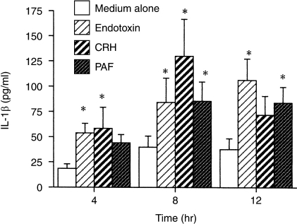
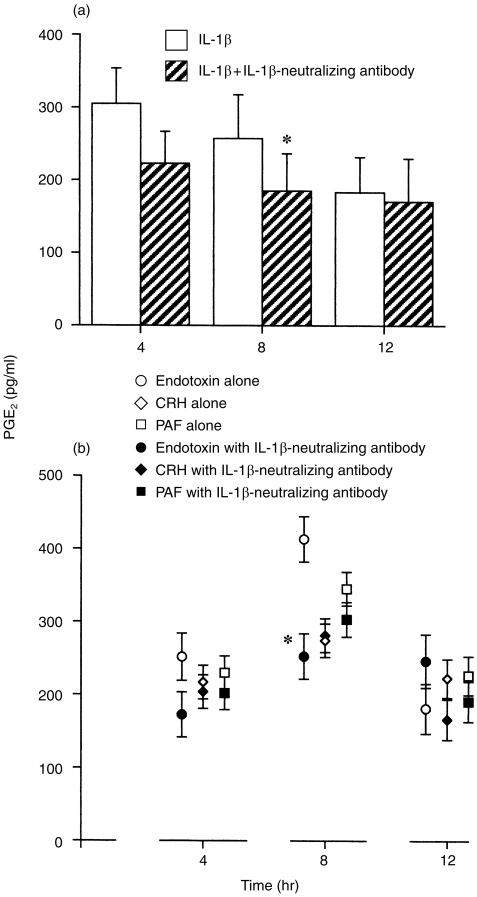
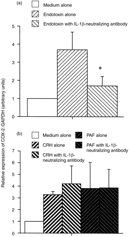
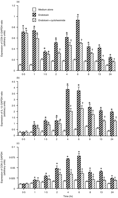
Bacterial endotoxin increased the expression of mRNA (maximal after 4 hr) for interleukin-1beta (IL-1beta) and the release of mature protein from intact human fetal membranes. In contrast, the change in expression of mRNA for type 2 cyclo-oxygenase (COX-2) was biphasic, with peaks after 0.5-1 hr and after 8 hr of culture. An antibody to IL-1beta was without effect after 4 hr of culture, inhibited endotoxin-stimulated prostaglandin E2 (PGE2) production after 8 hr of culture, and caused a parallel decrease in the expression of mRNA for COX-2. We conclude that endotoxin induced the expression of COX-2 through IL-1beta-independent and IL-1beta-dependent mechanisms, and these differences are time dependent. Corticotrophin-releasing hormone (CRH) or platelet-activating factor (PAF) also increased the expression of mRNA for IL-1beta and the release of IL-1beta from some, but not all, fetal membranes. The antibody to IL-1beta did not affect CRH-stimulated or PAF-stimulated PGE2 production or COX-2 expression. We conclude that CRH and PAF can induce the expression of IL-1beta, but this is not obligatory for increased PGE2 release, and the effect of these stimuli on COX-2 expression is a direct, IL-1beta-independent effect.
Figures

References
-
- Green K, Bygdeman M, Toppozada M, Wiqvist N. The role of prostaglandin F2α in human parturition. Am J Obstet Gynecol. 1974;120:25. - PubMed
-
- Lorenz RP, Botti JJ, Chez RA, Bennett N. Variations of biologic activity of low-dose prostaglandin E2 on cervical ripening. Obstet Gynecol. 1984;64:123. - PubMed
-
- Okazaki T, Casey ML, Okita JR, MacDonald PC, Johnston JM. Initiation of human parturition. XII. Biosynthesis and metabolism of prostaglandins in human fetal membranes and uterine decidua. Am J Obstet Gynecol. 1981;139:373. - PubMed
-
- Keirse MJNC, Turnbull AC. The fetal membranes as a possible source of amniotic fluid prostaglandins. Br J Obstet. 1976;83:146. - PubMed
-
- Khan H, Sullivan MHF, Helmig R, Roseblade CK, Uldbjerg N, Elder MG. Quantitative production of prostaglandin E2 and its metabolites by human fetal membranes. Br J Obstet Gynaecol. 1991;98:712. - PubMed
Publication types
MeSH terms
Substances
LinkOut - more resources
Full Text Sources
Research Materials
