. 1999 Jun;97(2):264-71.
doi: 10.1046/j.1365-2567.1999.00762.x.

Autoreactive cytotoxic T cells in mice are induced by immunization with a conserved mitochondrial enzyme in Freund's complete adjuvant

Affiliations

Autoreactive cytotoxic T cells in mice are induced by immunization with a conserved mitochondrial enzyme in Freund's complete adjuvant

V Karanikas et al. Immunology. 1999 Jun.

Abstract

Standard methods to generate autoimmune reactions in mice, by immunization with antigens emulsified with adjuvants, stimulate strong helper (CD4) T-cell and antibody responses but are not reported to induce cytolytic CD8 T cells. The aim of this study was to assess whether specific autoreactive CD8 T cells could be readily generated after immunization with a 'weak' autoantigen in adjuvant. Mice were immunized intraperitoneally three times with the E3 subunit of the mitochondrial 2-oxoacid dehydrogenase enzyme complexes (dihydrolipoamide dehydrogenase) emulsified with Freund's complete adjuvant. Splenic and lymph node lymphocytes were harvested after 14 days for in vitro functional studies. T lymphocytes were tested for proliferative responses and cytotoxicity against antigen-loaded isogeneic target cells. An autoreactive cytolytic T lymphocyte (CTL) response was detectable only after the in vitro restimulation of lymphocytes with E3 antigen-loaded syngeneic splenocytes. These CTL were identified as H-2-restricted CD8+ T cells. A proliferative response to E3 was demonstrable against antigen-pulsed syngeneic splenocytes. Immunized mice also generated strong antibody responses to E3. Liver histology showed portal infiltrates interpreted as a response of the liver to a non-specific immunological stimulus. It is concluded that autoreactive cytolytic T cells can be generated experimentally upon appropriate stimulation of the immune system, and can be identified in vitro upon release from the controlling mechanisms that are likely to regulate them in vivo.


Figures

Figure 1
Figure 1
Cytolytic responses after immunization of mice with PDC-E3 in FCA (n = 10) or PBS in FCA (n = 5). Effector cells were stimulated with irradiated syngeneic cells exposed to hypertonic medium and PDC-E3, and tested for lysis of isogeneic P1.PYT target cells (P1) or cells exposed to hypertonic medium and PDC-E3 (P1 + E3). The mean %SCR at E:T 40:1 are shown as horizontal bars, and the mean + 2SD &SCR value for cells from control mice immunized with PBS in FCA (dashed line) was used as a baseline to define positive responses.
Figure 2
Figure 2
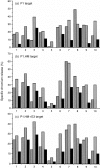
Dependence on in vitro antigen stimulation of cytolytic responses after immunization of mice with PDC-E3. Spleen cells from 10 mice (x-axis) were stimulated in vitro by either irradiated syngeneic lymphoid cells incubated with PDC-E3 in suspension (+ PDC-E3) (gray histogram), by syngeneic lymphoid cells treated with hypertonic medium to load PDC-E3 (HM/PDC-E3) (cross-hatched histograms), or by syngeneic lymphoid cells treated only with hypertonic medium (HM) (black histograms). After 7 days, responder cells from each culture were tested on three target cells: isogeneic P1.PYT target cells were untreated (a), treated with hypertonic medium only (b) or were loaded with PDC-E3 (c). Lysis at the E:T ratio 40:1 is shown.
Figure 3
Figure 3
H-2d-restricted CTL response from a mouse immunized with PDC-E3 in FCA. Effector cells were stimulated in vitro with irradiated syngeneic cells loaded with PDC-E3 and tested for lysis against either allogeneic L929 target cells (×) or isogeneic P1.PYT target cells that were untreated (•), exposed to hypertonic medium (▪), or exposed to hypertonic medium and loaded with PDC-E3 (▴).
Figure 4
Figure 4
Phenotype of cytolytic cells induced by immunization of mice with PDC-E3 in FCA. Lymphoid cells were stimulated in vitro with irradiated syngeneic cells loaded with PDC-E3 and prior to testing for cytolytic activity against isogeneic target cells, were incubated with either anti-CD4 or anti-CD8 monoclonal antibodies.
Figure 5
Figure 5
Immunization of mice with PDC-E3 in FCA induces a proliferative response. Lymphoid cells from eight mice given PDC-E3 in FCA (closed symbols) or three mice given PBS in FCA (open symbols) were stimulated in vitro with irradiated syngeneic spleen cells that were either: (i) untreated; (ii) exposed to PDC-E3 in suspension ( + PDC-E3); (iii) exposed to hypertonic medium (HM); or (iv) exposed to hypertonic medium containing PDC-E3 (HM/PDC-E3). Data (bars show mean±SD) are expressed as the ratio of c.p.m. for cells from mice given PDC-E3 in FCA to c.p.m. for cells from control untreated mice. Each data point is the mean of eight replicate wells.
Figure 6
Figure 6
Serum antibody to PDC-E3 measured by ELISA. Sera were tested at a dilution of 1:100 from five mice immunized with PBS in FCA (FCA/PBS) and 10 mice immunized with PDC-E3 in FCA (FCA/PDC-E3). The mean optical density at 415 nm is shown as a horizontal bar.
Figure 7
Figure 7
Histology of livers from BALB/c mice. Portal regions (arrowheads) of representative liver sections from examination of multiple mice: (a) a non-immunized mouse showing no portal cellular infiltration (H & E, ×270); (b) a mouse injected with PBS and FCA showing moderate ( + +) portal infiltration and an abnormal liver architecture, with the apparent sporadic death of hepatocytes and presence of lipid-like droplets (H & E, ×200); and (c) a mouse injected with PDC-E3 in FCA showing extensive and heavy (+ + +) portal infiltration (H & E, ×250). The internal scale marker represents 100 μm.

Similar articles

Cited by

References

    1. Bernard CCA, Mandel TE, MacKay IR. Experimental models of human autoimmune disease: Overview and prototypes. In: Rose NR, Mackay IR, editors. The Autoimmune Diseases II. Orlando: Academic Press; 1992. p. 47.
    1. Nagata M, Santamaria P, Kawamura T, Utsugi T, Yoon JW. Evidence for the role of CD8+ cytotoxic T cells in the destruction of pancreatic b cells in NOD mice. J Immunol. 1994;152:2042. - PubMed
    1. Yoneda R, Yokono K, Nagata M, et al. CD8 cytotoxic T-cell clone rapidly transfers autoimmune diabetes in very young NOD and MHC class I-compatible mice. Diabetologia. 1997;40:1044. - PubMed
    1. Kagi D, Odermatt B, Seiler P, Zinkernagel RM, Mak TW, Hengartner H. Reduced incidence and delayed onset of diabetes in perforin-deficient nonobese diabetic mice. J Exp Med. 1997;186:989. - PMC - PubMed
    1. Ohno S. ‘Self’ to cytotoxic T cells has to be 1000 or less high affinity nonapeptides per MHC antigen. Immunogenetics. 1992;36:22. - PubMed

Substances

LinkOut - more resources