

Human parathymic lymph node: morphological and functional significance
- PMID: 10447746
- PMCID: PMC2326821
- DOI: 10.1046/j.1365-2567.1999.00793.x
Human parathymic lymph node: morphological and functional significance
Abstract
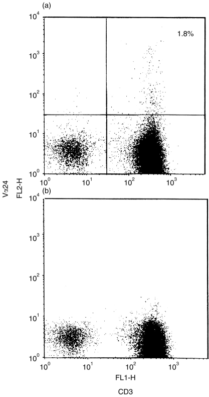
Parathymic lymph nodes (PTLNs) have been identified in several species, but in humans they have been noted only once before in a study 90 years ago using fetal material. We now report their occurrence in children. Human PTLNs are small but distinctive lymphatic organs located on the surface of the thymus (or sometimes between the upper and lower lobes of the thymus) and covered with the thymic capsule. Histologically, the medullary cords of these lymph nodes were found to be thin, with only small numbers of plasma cells. In addition, they had a well-developed paracortical area rich with high endothelial venules (HEV), but a thin cortex, including only a few undeveloped follicles. Flow cytometric analysis of PTLNs revealed that the ratios of T:B cells (14.6+/-9.3) and of CD4+:CD8+ T cells (4.9+/-1.4) in PTLNs were much higher than in other peripheral lymphoid tissues and in peripheral blood. Because of these characteristics of the human PTLNs, we propose that the human PTLNs might influence the functional differentiation of T cells.
Figures


Similar articles
-
Analyses of leucocytes in blood and lymphoid tissues from mink infected with Aleutian mink disease parvovirus (AMDV).Vet Immunol Immunopathol. 1998 Jun 12;63(4):317-34. doi: 10.1016/s0165-2427(98)00110-x. Vet Immunol Immunopathol. 1998. PMID: 9656422
-
Seeding of neonatal lymph nodes by T cells and identification of a novel population of CD3-CD4+ cells.Eur J Immunol. 1992 Feb;22(2):329-34. doi: 10.1002/eji.1830220207. Eur J Immunol. 1992. PMID: 1347010
-
IgE-bearing cells and epsilon-specific mRNA in lymphoid organs of two children with AIDS.Pediatr AIDS HIV Infect. 1997 Apr;8(2):102-7. Pediatr AIDS HIV Infect. 1997. PMID: 11361775
-
[Preliminary study on lymphocyte subsets of sentinel lymph nodes in breast cancer patients].Zhonghua Zhong Liu Za Zhi. 2004 Apr;26(4):220-2. Zhonghua Zhong Liu Za Zhi. 2004. PMID: 15312384 Chinese.
-
Role of parathymic lymph nodes in metastatic tumor development.Cancer Metastasis Rev. 2012 Jun;31(1-2):89-97. doi: 10.1007/s10555-011-9331-y. Cancer Metastasis Rev. 2012. PMID: 22134656 Review.
Cited by
-
Metastatic Spread from Abdominal Tumor Cells to Parathymic Lymph Nodes.Pathol Oncol Res. 2019 Apr;25(2):625-633. doi: 10.1007/s12253-018-0492-7. Epub 2018 Nov 7. Pathol Oncol Res. 2019. PMID: 30406399
-
[Morphology and functional anatomy of the growing thorax].Radiologe. 2003 Dec;43(12):1036-44. doi: 10.1007/s00117-003-0985-5. Radiologe. 2003. PMID: 14668991 Review. German.
References
-
- Severeanu G. Die Lymphgefäße der Thymus. Arch Anat Entw Gesch. 1909:93.
-
- Yamashita A, Miyasaka M, Trnka Z. Early post-thymic T cells: studies on lymphocytes in the lymph coming from thymus of the sheep. In: Morris B, Miyasaka M, editors. Immunology of the Sheep. Basel: Editions Roche; 1985. p. 162.
-
- Harris PF, Templeton WR. Preliminary studies on the lymphatic drainage of the guinea-pig thymus with special reference to extrinsic vessels. J Anat Lond. 1966;100:694.
-
- Harris PF, Templeton WR. Studies on the extrinsic lymphatic drainage of the guinea-pig thymus. Acta Anat. 1968;69:366. - PubMed
Publication types
MeSH terms
Substances
LinkOut - more resources
Full Text Sources
Research Materials
