. 1999 Jul;97(3):420-8.
doi: 10.1046/j.1365-2567.1999.00803.x.

Enterobacterial infection modulates major histocompatibility complex class I expression on mononuclear cells

Affiliations

Enterobacterial infection modulates major histocompatibility complex class I expression on mononuclear cells

J Kirveskari et al. Immunology. 1999 Jul.

Abstract

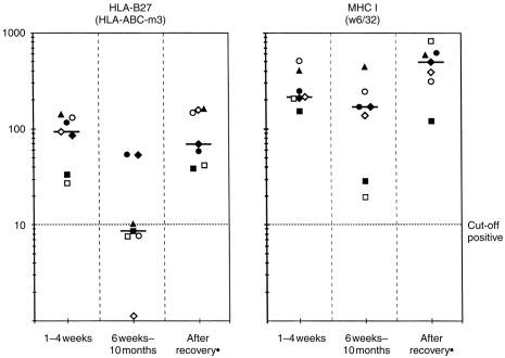
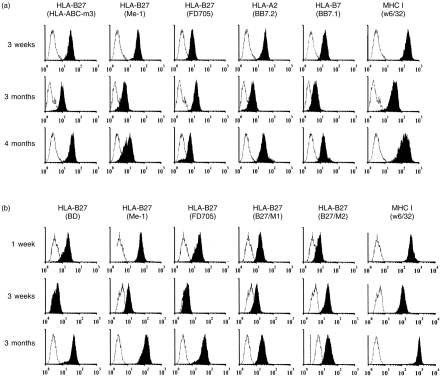
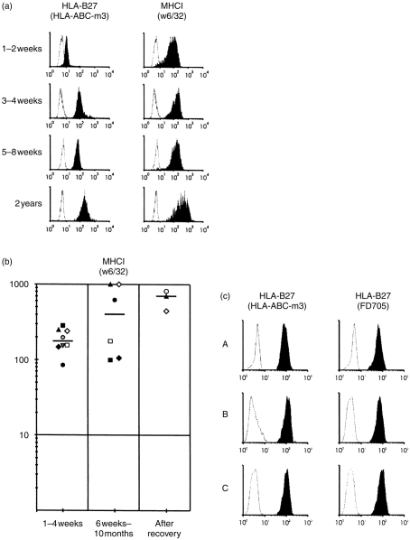
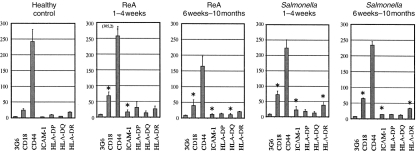
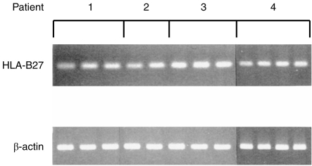
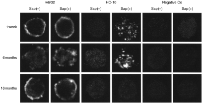
Major histocompatibility complex (MHC) class I expression is reduced in several viral infections, but it is not known whether the same happens during infections caused by intracellular enterobacteria. In this study, the expression of MHC class I antigens on peripheral blood mononuclear cells (PBMC) from 16 patients with Salmonella, Yersinia, or Klebsiella infection was investigated. During or after the acute infection, the expression of MHC class I antigens was markedly decreased in eight patients, all with genotype HLA-B27, and six out of eight with reactive arthritis (ReA). A significant decrease of monomorphic MHC class I was found in three patients, of HLA-B27 in eight (P<0.05) and of HLA-A2 in two. However, patients negative for the HLA-B27 genotype, or healthy HLA-B27-positive individuals, did not have a significant decrease of MHC class I antigens. During the decreased expression on the cell surface, intracellular retention of MHC class I antigens was observed, whereas HLA-B27 mRNA levels did not vary significantly. This is the first evidence that enterobacterial infection may down-regulate expression of MHC class I molecules in vivo and that down-regulation is predominant in patients with the HLA-B27 genotype.


Figures

Figure 1
Figure 1
Expression of human leucocyte antigen (HLA)-B27 (a) and monomorphic major histocompatibility complex (MHC) class I (b) molecules on monocytes from seven patients with Salmonella-triggered reactive arthritis (ReA) who were positive for the HLA-B27 gene during the acute phase of infection (1–4 weeks, patient 3 after 8 weeks) and during the follow-up. *Patients ◊ and ▪ did not recover during the follow-up. The cells were incubated with monoclonal antibodies (mAbs) specific for HLA-B27 (HLA-ABC-m3) and monomorphic MHC class I (w6/32) and then with fluorescein isothiocyanate (FITC)-labelled F(ab′)2 fragments of goat antimouse IgG. The y-axis is the mean fluorescence intensity (MFI) of the specific mAb. The MFI of the negative control mAb was subtracted from each value before plotting.
Figure 2
Figure 2
(a) Expression of human leucocyte antigen (HLA)-B27, HLA-A2, HLA-B7 and major histocompatibility complex (MHC) class I molecules on monocytes from patient 5 (P5) with Salmonella-triggered reactive arthritis (ReA) at the start of the infection and during the follow-up. The cells were incubated with monoclonal antibodies (mAbs) specific for HLA-B27 (three mAbs against different epitopes of HLA-B27), HLA-A2, HLA-B7 and monomorphic MHC class I and then with fluorescein isothiocyanate (FITC)-labelled F(ab′)2 fragments of goat antimouse IgG. The expression of MHC class I epitopes recognized with mAbs is shown as black histograms and corresponding negative controls as white histograms. The x-axis is the mean fluorescence intensity (MFI) on a log scale, and the y-axis is the relative number of the cells. (b) Expression of HLA-B27 and monomorphic MHC class I epitopes on monocytes from a patient with Yersinia-triggered ReA. The cells were incubated with mAbs specific for HLA-B27 (four mAbs against different epitopes of HLA-B27) and monomorphic MHC class I and then with FITC-labelled F(ab′)2 fragments of goat antimouse IgG.
Figure 3
Figure 3
(a) Expression of human leucocyte antigen (HLA)-B27 and major histocompatibility complex (MHC) class I molecules on monocytes from the patient with uncomplicated Salmonella infection. The cells were incubated with monoclonal antibodies (mAbs) specific for HLA-B27 or with negative control mAbs, and then with fluorescein isothiocyanate (FITC)-labelled F(ab′)2 fragments of goat antimouse IgG. The expression of HLA-B27 detected with mAb HLA-ABC-m3 is shown as black histograms and corresponding negative controls as white histograms. The x-axis is the mean fluorescence intensity (MFI) on a log scale and the y-axis is the relative number of the cells. (b) Expression of monomorphic MHC class I on monocytes from patients with S. enteritidis infection without reactive arthritis (ReA). The cells were incubated with mAb recognizing monomorphic MHC class I and then with FITC-labelled F(ab′)2 fragments of goat antimouse IgG. The y-axis is the MFI of the specific mAb. The MFI of the negative control mAb was subtracted from each value before plotting. (c) Expression of HLA-ABC-m3 and FD705 epitopes of HLA-B27 on monocytes from a healthy control subject (A, B and C). The second sample (B) was taken 5 weeks and the third sample (C) 1 year after the first sample (A).
Figure 4
Figure 4
Expression of adhesion/co-stimulatory and major histocompatibility complex (MHC) class II molecules on peripheral blood mononuclear cells (PBMC) during the acute phase of Salmonella infection and during the follow-up. Shown are the mean fluorescence intensities (MFI) (±SEM). The control group consisted of seven healthy blood donors. The non-specific background staining with the appropriate negative control monoclonal antibody (mAb) was subtracted from the staining with the given mAb. Statistical significance was evaluated using the Mann–Whitney U-test. *P < 0·05; **P < 0·005. The MFI data in this figure from seven healthy control subjects and four patients with uncomplicated Salmonella infection has been published previously.17
Figure 5
Figure 5
Expression of human leucocyte antigen (HLA)-B27 mRNA in the follow-up samples of four patients (P1, P2, P3 and P7; from P2 only the first two samples are shown). Total RNA was isolated from peripheral blood mononuclear cells (PBMC) of the patients, and the expression of HLA-B27 mRNA was analysed using semiquantitative reverse transcription–polymerase chain reaction (RT–PCR).
Figure 6
Figure 6
Expression of major histocompatibility complex (MHC) class I antigens on peripheral blood mononuclear cells (PBMC) of a patient with Salmonella-triggered reactive arthritis (ReA). The cells were incubated with a monoclonal antibody (mAb) recognizing monomorphic MHC class I antigens (w6/32), free heavy chain (HC-10), or with a negative control (3G6), and then with fluorescein isothiocyanate (FITC)-labelled F(ab′)2 fragments of goat antimouse IgG. The PBMC were either stained for cell surface antigens (Sap(−)) or permeabilized with saponin (Sap(+)) for intracellular staining. The PBMC were analysed by confocal microscopy. The middle sections of representative cells are shown.

Similar articles

Cited by

References

    1. Garrido F, Ruiz-Cabello F, Cabrera T, et al. Implications for immunosurveillance of altered HLA class I phenotypes in human tumours. Immunol Today. 1997;18:89. - PubMed
    1. Andersson M, Pääbo S, Nilsson T, et al. Impaired intracellular transport of class I MHC antigens as a possible means for adenoviruses to evade immune surveillance. Cell. 1985;43:215. - PubMed
    1. Hill AB, Barnett BC, McMichael AJ, et al. HLA class I molecules are not transported to the cell surface in cells infected with herpes simplex virus types 1 and 2. J Immunol. 1994;152:2736. - PubMed
    1. Ploegh HL. Viral strategies of immune evasion. Science. 1998;280:248. - PubMed
    1. Sieper J, Braun J. Pathogenesis of spondylarthropathies. Persistent antigen, autoimmunity, or both? Arthritis Rheum. 1995;38:1547. - PubMed

Publication types

MeSH terms