Platelet-activating factor receptor mRNA is localized in eosinophils and epithelial cells in rat small intestine: regulation by dexamethasone and gut flora
- PMID: 10447766
- PMCID: PMC2326856
- DOI: 10.1046/j.1365-2567.1999.00784.x
Platelet-activating factor receptor mRNA is localized in eosinophils and epithelial cells in rat small intestine: regulation by dexamethasone and gut flora
Abstract
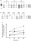
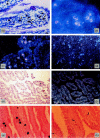
Platelet-activating factor (PAF) is a potent mediator involved in bowel injury. We investigated PAF receptor transcription and its mRNA localization in the small intestine of normal (conventionally fed) and germ-free rats, by competitive polymerase chain reaction (PCR) and in situ hybridization. A dose of PAF (1.5 microg/kg, i.v.) insufficient to cause gross bowel injury was injected into rats. Some rats were pretreated with dexamethasone (1 mg/kg). We found: (1) PAF receptor (PAF-R) mRNA localized predominantly in lamina propria eosinophils and in epithelial cells; (2) PAF increased PAF-receptor signals in the epithelial cells; (3) Dexamethasone depleted eosinophils in the intestine and markedly decreased PAF-receptor transcripts; the response to PAF was also weaker than control rats; (4) Germ-free rats had less PAF-R mRNA than normal rats, and showed a weaker response to PAF than conventionally fed rats. Thus, we conclude: (1) PAF receptor mRNA is constitutively expressed in the epithelium and in lamina propria eosinophils in the intestine. (2) PAF-R transcription is up-regulated by PAF and gut flora, mostly in the epithelium. (3) PAF-R transcription is down-regulated by glucocorticoids, mainly as a result of eosinophil depletion. These results suggest a functional role for PAF receptors both in host defence and the inflammatory response in the small intestine.
Figures


Similar articles
-
Human intestinal epithelial cells express receptors for platelet-activating factor.Am J Physiol. 1999 Oct;277(4):G810-8. doi: 10.1152/ajpgi.1999.277.4.G810. Am J Physiol. 1999. PMID: 10516147
-
Regulation of platelet-activating factor receptor gene expression in vivo by endotoxin, platelet-activating factor and endogenous tumour necrosis factor.Biochem J. 1997 Mar 1;322 ( Pt 2)(Pt 2):603-8. doi: 10.1042/bj3220603. Biochem J. 1997. PMID: 9065783 Free PMC article.
-
Increased platelet-activating factor receptor gene expression by corneal epithelial wound healing.Invest Ophthalmol Vis Sci. 2000 Jun;41(7):1696-702. Invest Ophthalmol Vis Sci. 2000. PMID: 10845588
-
Platelet activating factor/platelet activating factor receptor pathway as a potential therapeutic target in autoimmune diseases.Inflamm Allergy Drug Targets. 2009 Jul;8(3):182-90. doi: 10.2174/187152809788681010. Inflamm Allergy Drug Targets. 2009. PMID: 19601878 Review.
-
Platelet-activating factor receptor: gene expression and signal transduction.Biochim Biophys Acta. 1995 Dec 7;1259(3):317-33. doi: 10.1016/0005-2760(95)00171-9. Biochim Biophys Acta. 1995. PMID: 8541341 Review. No abstract available.
Cited by
-
The Eosinophil in Health and Disease: from Bench to Bedside and Back.Clin Rev Allergy Immunol. 2016 Apr;50(2):125-39. doi: 10.1007/s12016-015-8507-6. Clin Rev Allergy Immunol. 2016. PMID: 26410377 Review.
-
Identification of Differential Protein Expression in Hepatocellular Carcinoma Induced Wistar Albino Rats by 2D Electrophoresis and MALDI-TOF-MS Analysis.Indian J Clin Biochem. 2016 Apr;31(2):194-202. doi: 10.1007/s12291-015-0510-4. Epub 2015 May 26. Indian J Clin Biochem. 2016. PMID: 27069327 Free PMC article.
-
Inflammatory signaling in necrotizing enterocolitis.Clin Perinatol. 2013 Mar;40(1):109-24. doi: 10.1016/j.clp.2012.12.008. Epub 2013 Jan 17. Clin Perinatol. 2013. PMID: 23415267 Free PMC article. Review.
-
Inflammatory signaling in NEC: Role of NF-κB, cytokines and other inflammatory mediators.Pathophysiology. 2014 Feb;21(1):55-65. doi: 10.1016/j.pathophys.2013.11.010. Epub 2013 Dec 31. Pathophysiology. 2014. PMID: 24388163 Free PMC article. No abstract available.
-
Platelet-activating factor increases mucosal permeability in rat intestine via tyrosine phosphorylation of E-cadherin.Br J Pharmacol. 2000 Apr;129(7):1522-9. doi: 10.1038/sj.bjp.0702939. Br J Pharmacol. 2000. PMID: 10742310 Free PMC article.
References
-
- Benveniste J. Paf-acether, an ether phospho-lipid with biological activity. Prog Clin Biol Res. 1988;282:73. - PubMed
-
- Nakamura M, Honda Z, Izumi T, et al. Molecular cloning and expression of platelet-activating factor receptor from human leukocytes. J Biol Chem. 1991;266:0400. - PubMed
-
- Kishimoto S, Shimadzu W, Izumi T, et al. Regulation by IL-5 of expression of functional platelet-activating factor receptors on human eosinophils. J Immunol. 1996;157:4126. - PubMed
-
- Franklin RA, Mazer B, Sawami H, et al. Platelet-activating factor triggers the phosphorylation and activation of MAP-2 kinase and S6 peptide kinase activity in human B cell lines. J Immunol. 1993;151:1802. - PubMed
Publication types
MeSH terms
Substances
Grants and funding
LinkOut - more resources
Full Text Sources
Molecular Biology Databases

