. 1999 Jul;97(3):466-73.
doi: 10.1046/j.1365-2567.1999.00778.x.

Intravenous tolerization with type II collagen induces interleukin-4-and interleukin-10-producing CD4+ T cells

Affiliations

Intravenous tolerization with type II collagen induces interleukin-4-and interleukin-10-producing CD4+ T cells

M L Gumanovskaya et al. Immunology. 1999 Jul.

Abstract

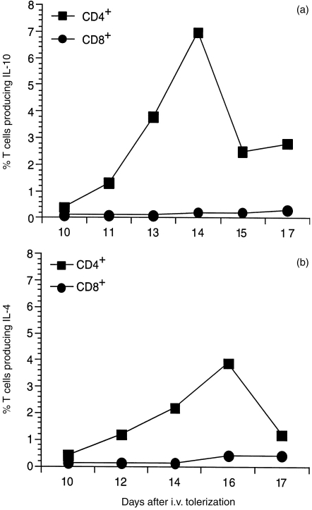
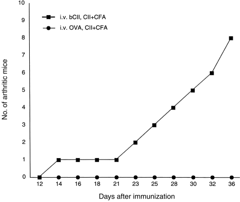
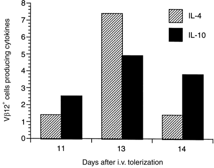
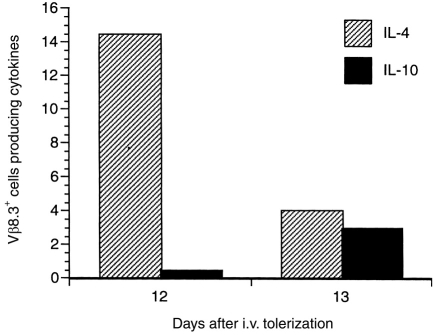
Intravenous (i.v.) administration of type II collagen (CII) is an effective way to induce tolerance and suppress disease in the collagen-induced arthritis (CIA) model. In this study, we demonstrated that a single i.v. dose of CII (as low as 0.1 mg/mouse) completely prevented the development of CIA. This suppression was accompanied by decreases in levels of antibody specific for the immunogen, bovine CII and autoantigen, mouse CII. Splenocytes obtained from CII-tolerized mice and stimulated with CII in vitro produced predominantly the T helper 2 (Th2)-type cytokines interleukin-4 (IL-4) and interleukin-10 (IL-10). In contrast, cells obtained from mice immunized with CII produced predominantly interferon-gamma (IFN-gamma). Two-colour flow cytometric analysis of cytokine expression and T-cell phenotype demonstrated that CD4+ cells and not CD8+ or gammadelta+ cells were the predominant regulatory cells producing IL-4 and IL-10. Transgenic mice bearing a T-cell receptor (TCR) specific for CII had a greater increase in the number of IL-4-secreting CD4+ cells, as well as a marked increase of IL-4 in culture supernatants. This cytokine was produced by transgene-bearing T cells. Elucidation of mechanisms for the induction of tolerance in mature T cells is an important line of study in autoimmune models because of the potential application for treating organ-specific autoimmune disease.


Figures

Figure 1
Figure 1
Splenocytes from DBA/1 mice tolerized intravenously with 1 mg of type II collagen (CII) were obtained at intervals, stimulated in vitro with bovine α1(II) for 8 hr and stained with fluorescein isothiocyanate (FITC)-conjugated antibody specific for CD4 or CD8. Cells were counter-stained with phycoerythrin (PE)-conjugated anticytokine monoclonal antibody (mAb) recognizing interleukin (IL)-4 or IL-10 and analysed on an EPICS Profile II cytofluorometer. The quadrant markers of the bivariant dot were set based on negative staining controls using isotype-matched immunoglobulin controls. IL-10 (a) was secreted by CD4+ T cells and peaked at day 14 after tolerization; IL-4 (b) was also secreted by CD4+ cells and peaked at day 16.
Figure 2
Figure 2
Vβ8.3tg mice were injected intravenously (i.v.) with either ovalbumin (OVA; negative control) or bovine type II collagen (CII). Protein (333 μg) was administered daily for 3 days so that each mouse received a total of 1000 μg of either protein. Mice were immunized with CII emulsified in complete Freund’s adjuvant (CFA) 4 days after the last i.v. dose and mice were observed for the incidence of arthritis.
Figure 3
Figure 3
Spleen cells, from SWRβTg mice that had been administered intravenously (i.v.) 1 mg of type II collagen (CII), were obtained at intervals after i.v. injection and stimulated with bovine α1(II) CII in vitro as described in the Materials and methods. Two-colour surface receptor and intracellular cytokine staining were performed. Data represent the proportion of the dual-positive cells for Vβ12 and cytokine markers among mononuclear cells.
Figure 4
Figure 4
Spleen cells, from DBAVβ8.3Tg mice that had been administered intravenously (i.v.) 1 mg of type II collagen (CII), were obtained at intervals after i.v. injection and stimulated with bovine α1(II) CII in vitro as described in the Materials and Methods. Two-colour surface receptor and intracellular cytokine staining were performed. Data represent the proportion of the dual-positive cells for Vβ8.3 and cytokine markers among mononuclear cells.

Similar articles

Cited by

References

    1. Cremer MA, Hernandez AD, Stuart JM, Townes AS, Kang AH. Collagen-induced arthritis in rats: antigen specific suppression of arthritis and immunity by intravenously injected native type II collagen. J Immunol. 1983;131:2995. - PubMed
    1. Nagler-Anderson CN, Bober LA, Robinson ME, Siskind GW, Thorbecke GJ. Suppression of type II collagen-induced arthritis by intragastric administration of soluble type II collagen. Proc Natl Acad Sci USA. 1986;83:7443. - PMC - PubMed
    1. Thompson HS, Staines NA. Gastric administration of type II collagen delays the onset and severity of collagen-induced arthritis in rats. Clin Exp Immunol. 1986;64:581. - PMC - PubMed
    1. Wooley PH, Luthra HS, Singh S, Huse A, Stuart JM, David CS. Passive transfer of arthritis in mice by human anti-type II collagen antibody. Mayo Clinic Proc. 1984;59:737. - PubMed
    1. Myers L, Seyer J, Stuart J, Kang A. Suppression of murine collagen-induced arthritis by nasal administration of collagen. Immunology. 1997;90:161. - PMC - PubMed

Publication types

LinkOut - more resources