. 1999 Jun;2(6):528-34.
doi: 10.1038/9183.

Target-specific control of nicotinic receptor expression at developing interneuronal synapses in chick

Affiliations

Target-specific control of nicotinic receptor expression at developing interneuronal synapses in chick

P Devay et al. Nat Neurosci. 1999 Jun.

Abstract

Neuronal differentiation and development of synaptic specializations are strongly influenced by cellular interactions. We compared the effects of interaction with distinct autonomic targets on the molecular and biophysical differentiation of 'upstream' neuron-neuron synapses. Contact with cardiac tissue induced expression of nicotinic receptor channels (nAChRs) distinct from those induced by renal tissue in presynaptic autonomic neurons. The kinetics of cholinergic currents at interneuronal synapses are dictated by the peripheral target contacted. Analysis of the nAChR channel subtypes and subunits in individual neurons demonstrated that the profile of transmitter receptors expressed at mature neuron-neuron synapses develops from the convergent influences of input-derived (anterograde) and target-specific (retrograde) signals.


Figures
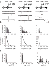
Fig. 1
Fig. 1
Spontaneous synaptic currents from individual innervated sympathetic neurons: effect of target interactions. (a) Schematic diagram of cellular interactions and recording configurations (above); sample recordings from three innervated sympathetic neurons maintained in vitro in the presence or absence of the indicated targets (below). Synaptic currents were recorded from (left to right) an innervated sympathetic neuron in the absence of target tissue (input + SNs), an innervated sympathetic neuron contacting renal tissue (input + SNs + kidney) and an innervated sympathetic neuron contacting cardiac tissue (input + SNs + heart). Scale bar, 40 pA, 40 ms. (b) Histograms of the sEPSC amplitudes from the same three neurons (above). The sEPSC amplitude distributions are skewed, precluding the use of mean amplitude as a representative measure of sEPSC size. Cumulative amplitude distributions of the sEPSCs recorded from the same three neurons (below). The area of each distribution is calculated to yield an ‘amplitude index’ for each cell (see Methods). Amplitude indices for these three cells are 24.3, 38.7 and 8.9 respectively. (c) Distributions of the decay time constants of the sEPSCs recorded from the same three neurons.
Fig. 2
Fig. 2
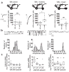
Target-specific regulation of nAChR-mediated macroscopic and synaptic currents. Effects of target contact on synaptic nAChRs were examined in innervated neurons without target (n = 33), contacting kidney (n = 29) and contacting heart (n = 31). (a) ACh-evoked macroscopic currents recorded in innervated sympathetic neurons without target or in those contacting either renal or cardiac target tissue. Responses of innervated neurons lacking target were similar to those of neurons contacting cardiac tissue; their distributions were unimodal with means of 949 ± 143 pA (χ2 = 0.6, n = 17) and 785 ± 53 (χ2 = 0.3, n = 13), respectively. In contrast, the distribution of ACh-elicited macroscopic currents of innervated neurons contacting renal tissue was bimodal; most responses were significantly larger than those recorded in innervated neurons with or without target contact (1760 ± 39 pA; n = 7 of 11, χ2 = 0.02). (b) sEPSC amplitude index values for innervated sympathetic neurons maintained without target (top) or with either renal (middle) or cardiac tissue (bottom) are shown. The amplitude index distribution for targetless, innervated neurons was well fit by the sum of two Gaussian distributions, with 87% of the values within the first peak centered at 20.1 ± 0.6 (χ2 = 0.44). The distribution of sEPSC amplitude indices for innervated neurons contacting kidney was multimodal, with 52% of the values in the first and 34% in the second Gaussian peak (at 15.9 ± 1.6 and at 35.8 ± 1.6 respectively; χ2= 1.04) and 14% of the values ≥ 50. In contrast, the distribution of the amplitude index values for innervated neurons contacting heart was described by a single log-normal distribution centered at 11.63 ± 0.83 (χ2 = 0.61). (c) Plots of the decay time constants (τsyn) of sEPSCs recorded in innervated neurons contacting kidney or heart or in the absence of target. The time constants of synaptic events recorded in targetless neurons and in neurons contacting kidney reflect uniformly fast nAChR kinetics (no target, τsyn = 2.08 ± 0.05; n = 1546, χ2= 2.8; +kidney, τsyn = 2.23 ± 0.03, n = 2258, χ2 = 2.8). In contrast, sEPSCs in most neurons contacting heart reflect roughly equal contributions of both fast and slow events (48%, τf = 2.09 ± 0.03 ms; 42%, τs1 = 16.9 ± 0.8 ms) with the slowest synaptic currents (τs2 = 37.1 ± 3 ms) making up 9% of the currents recorded (n = 699; χ2 = 0.9).
Fig. 3
Fig. 3
Synaptic nAChR channel properties and subunit gene expression are regulated by target. After electrophysiology (Fig. 2), levels of nAChR subunit mRNA of each neuron were measured by quantitative PCR. (a) Effects of renal or cardiac target on nAChR expression. Innervated neurons with or without kidney target are characterized by uniformly fast kinetic sEPSCs and by significantly higher α3 and α7 and lower β4 expression than innervated neurons with both fast and slow kinetic synaptic events (that is, those contacting heart). Relative expression levels of α3 to α5 + α7 + β4 in individual innervated neurons with fast sEPSCs (+ kidney) are compared with those with both fast and slow τsyn (+heart). Data were analyzed using non-parametric statistics and are presented as ‘box plots’ (see Methods). (b) Relationship between nAChR-subunit expression and sEPSC amplitude in innervated neurons with or without different targets. In innervated neurons either lacking target or contacting kidney, amplitude index values are positively correlated with ratio of α3/(α5 + β4); r = 0.93 and 0.97, respectively). In contrast, the ratio of α3 to α5 and β4 is low in neurons contacting heart, regardless of sEPSC amplitude index. Likewise, the ratio of α3/α5 is positively correlated with amplitude index in innervated neurons contacting kidney, whereas this ratio is constant over the full range of amplitudes in innervated neurons contacting heart. (c) Target regulation of nAChR subunit expression in innervated neurons. Innervated neurons contacting kidney have significantly higher ratios of α3 to α5 and/or to β4 than innervated neurons contacting cardiac tissue. *p ≤ 0.01; **p ≤ 0.002.
Fig. 4
Fig. 4
Target contact differentially regulates the profile of nAChR channels and subunit gene expression in non-innervated sympathetic neurons. (a) Schematic diagram of cellular interactions and recording configurations (above) and sample macroscopic currents elicited by 2.5 mM ACh (below) from non-innervated sympathetic neurons maintained in the absence of target (SNs alone), neurons contacting kidney (SNs + kidney) and neurons contacting heart (SNs + heart). Amplitudes of currents evoked by 500 μM ACh were significantly larger in neurons contacting renal tissue (n = 27) than those elicited in neurons maintained without target (n = 49, p < 0.001). In contrast, macroscopic currents recorded in neurons contacting cardiac tissue are significantly smaller than control (n = 19, p < 0.004). Increasing ACh concentration from 500 μM to 2.5 mM significantly increased current in neurons with kidney, but had little effect under other culture conditions. Data were analyzed using non-parametric statistics and are presented as box plots (see Methods). Note the difference in scale on amplitude axes. (b) Uninnervated sympathetic neurons express three nAChR channel subtypes classified on the basis of distinct chord conductances, pharmacology and kinetics,,. Representative traces from single-channel recordings in the three experimental groups (top). The peaks in the amplitude distributions from control neurons correspond to ~15, 30 and 50 pS nAChR channels (n = 11; bottom). Neurons contacting kidney express two major conductance classes with relatively brief opening times: one 30–35 pS and the other ≥ 70 pS (n = 25),,. In contrast, neurons contacting heart primarily express longer-duration nAChRs of ≲15 pS (n = 9). (c) The levels of nAChR subunit gene expression were measured in 30–40 neurons per experiment by qPCR of cytoplasmic RNA (see Methods and Fig. 5) and were plotted relative to actin mRNA levels. We assayed neurons contacting kidney (n = 8), contacting heart (n = 9) and without target (n = 15). Contact with kidney significantly increased α3, α5 and β4 from control (targetless) levels. In contrast, contact with cardiac tissue had little effect on α3 mRNA levels but significantly upregulated α5, α7 and β4 expression. *p ≤ 0.01; **p ≤ 0.001; ***p ≤ 0.0001.
Fig. 5
Fig. 5
Characterization of qPCR assay used to measure α3, α5, α7 and β4 mRNA levels in individual neurons. (a) Above, an agarose gel showing the amplified actin, α3, α5, α7 and β4 PCR products from a single innervated neuron. Below, agarose gel of a standard curve of PCR products amplified from decreasing amounts of subunit cDNAs. (b) Amplification of subunit mRNAs from a single neuron for 2 × 35 + 1 × 20 cycles yields DNA product within the linear range of standard curves generated with 0.05 to 1 fg of template. (c) Standard curve over the entire range. Amplified product was quantified based on [32P]dNTP incorporation.

Similar articles

Cited by

References

    1. Ozaki M, Sasner M, Yano R, Lu HS, Buonanno A. Neuregulin-beta induces expression of an NMDA-receptor subunit. Nature. 1997;390:691–694. - PubMed
    1. Yang X, Kuo Y, Devay P, Yu C, Role L. A cysteine-rich isoform of neuregulin controls nicotinic receptor expression in neurons during synaptogenesis. Neuron. 1998;20:255–270. - PubMed
    1. Devay P, Qu X, Role LW. Regulation of nAChR-subunit gene expression relative to the development of pre- and postsynaptic projections of embryonic chick sympathetic neurons. Dev Biol. 1994;162:56–70. - PubMed
    1. Moss BL, Schuetze SM, Role LW. Functional properties and developmental regulation of nicotinic acetylcholine receptors on embryonic chicken sympathetic neurons. Neuron. 1989;3:597–607. - PubMed
    1. Jacob MH, Berg DK. Effects of preganglionic denervation and postganglionic axotomy on acetylcholine receptors in the chick ciliary ganglion. J Cell Biol. 1987;105:1847–1854. - PMC - PubMed

Publication types