. 1999 Aug;104(4):399-407.
doi: 10.1172/JCI6629.

Ablation of the PTHrP gene or the PTH/PTHrP receptor gene leads to distinct abnormalities in bone development

Affiliations

Ablation of the PTHrP gene or the PTH/PTHrP receptor gene leads to distinct abnormalities in bone development

B Lanske et al. J Clin Invest. 1999 Aug.

Abstract

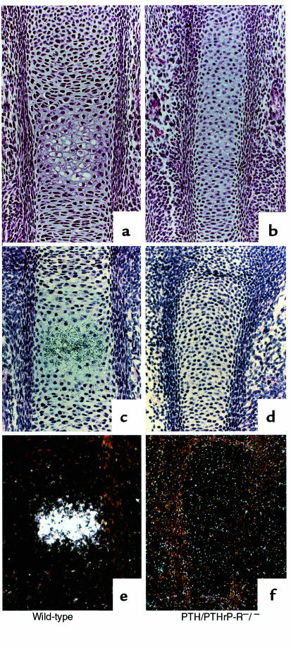
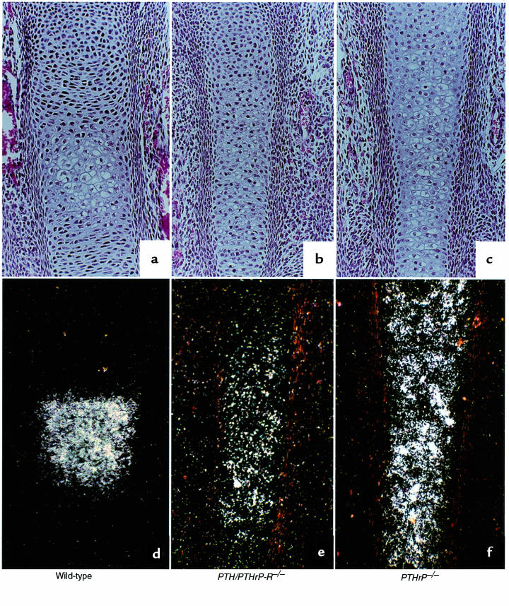
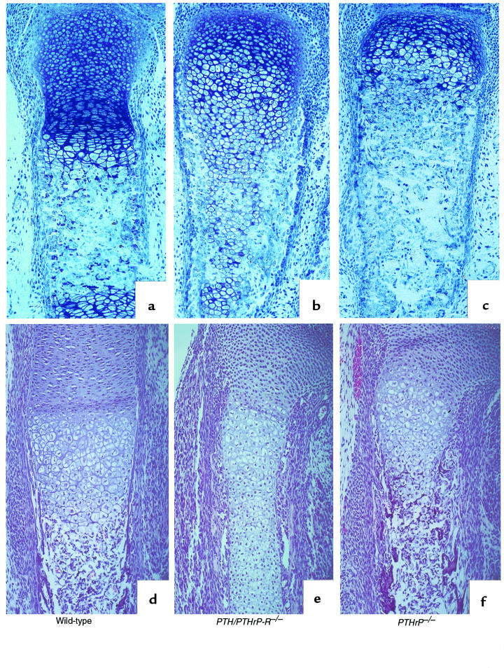
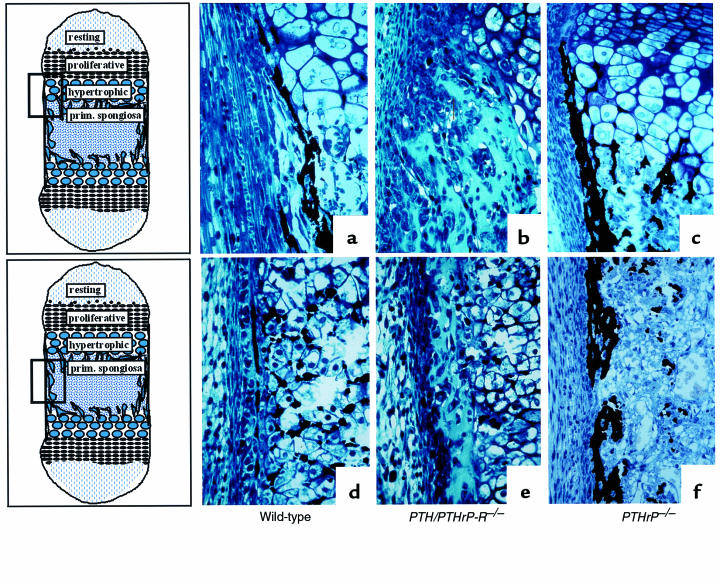
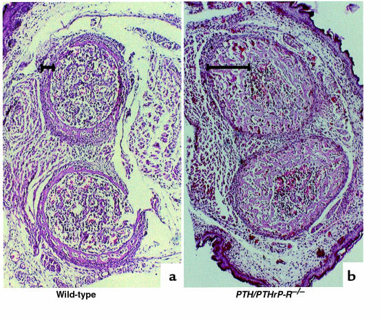
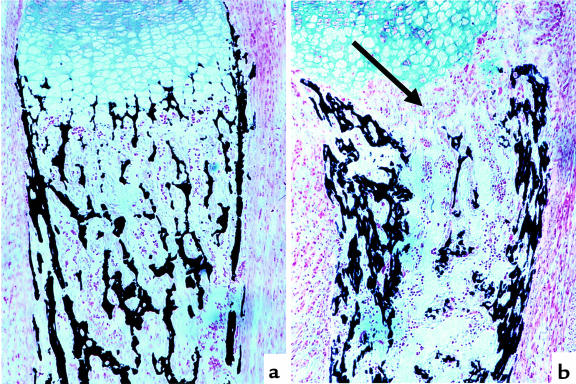
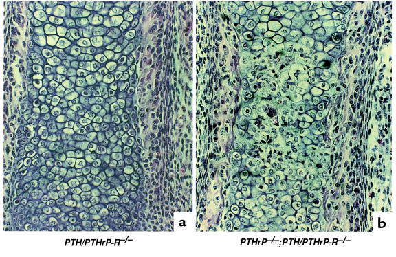
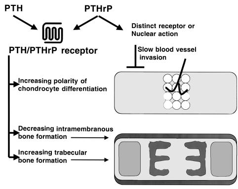
Parathyroid hormone (PTH) and parathyroid hormone-related peptide (PTHrP) bind to and activate the same PTH/PTHrP receptor. Deletion of either the PTHrP gene or the PTH/PTHrP receptor gene leads to acceleration of differentiation of growth plate chondrocytes. To explore further the functional relationships of PTHrP and the PTH/PTHrP receptor, bones of knockout mice were analyzed early in development, and the phenotypes of double-knockout mice were characterized. One early phenotype is shared by both knockouts. Normally, the first chondrocytes to become hypertrophic are located in the centers of long bones; this polarity is greatly diminished in both these knockouts. The PTH/PTHrP receptor-deficient (PTH/PTHrP-R(-/-)) mice exhibited 2 unique phenotypes not shared by the PTHrP(-/-) mice. During intramembranous bone formation in the shafts of long bones, only the PTH/PTHrP-R(-/-) bones exhibit a striking increase in osteoblast number and matrix accumulation. Furthermore, the PTH/PTHrP-R(-/-) mice showed a dramatic decrease in trabecular bone formation in the primary spongiosa and a delay in vascular invasion of the early cartilage model. In the double-homozygous knockout mice, the delay in vascular invasion did not occur. Thus, PTHrP must slow vascular invasion by a mechanism independent of the PTH/PTHrP receptor.


Figures

Figure 1
Figure 1
Chondrocyte differentiation at E15.5. Hematoxylin/eosin staining (a and b) and type X collagen mRNA in situ hybridization (cf) of wild-type and PTH/PTHrP-R–/– phalanges. PTH/PTHrP receptor–ablated bones show a delay in chondrocyte differentiation and type X collagen expression (b, d, and f) when compared with wild-type bones (a, c, and e). Bright-field, c and d; dark-field, e and f.
Figure 2
Figure 2
Chondrocyte differentiation at E16.5. Hematoxylin/eosin staining (ac) and type X collagen mRNA in situ hybridization (df) of wild-type, PTH/PTHrP-R–/–, and PTHrP–/– phalanges at E16.5. In contrast to PTHrP–/– bones, which exhibit a spatial and temporal progression in chondrocyte differentiation (c and f), PTH/PTHrP-R–/– bones show a delay in chondrocyte differentiation, as shown by the decrease in type X collagen expression (b and e), when compared with wild-type bones (a and d). However, the spatial distribution of the abnormally differentiated hypertrophic cells is identical (b, c and e, f).
Figure 3
Figure 3
Delay in replacement of cartilage by bone. Methylmethacrylate sections of phalanges at E18.5 (ac) and paraffin sections of tibiae at E16.5 (df) of wild-type, PTH/PTHrP-R–/–, and PTHrP–/– animals. Blood vessel invasion and deposition of bone by osteoblasts are delayed in PTH/PTHrP-R–/– bones (b and e) when compared with wild-type bones (a and d). In contrast, in PTHrP–/– bones, these processes are advanced (c and f).
Figure 4
Figure 4
Increase in osteoblast number and cortical bone in PTH/PTHrP-R–/– animals. Von Kossa staining on methylmethacrylate sections at the level of the metaphyseal region of a tibia (ac) and at the diaphyseal region of a phalanx (df) in wild-type, PTHrP–/–;PTH/PTHrP-R–/–, and PTHrP–/– animals at E18.5. PTH/PTHrP receptor mutant bones reveal an abnormal augmentation in osteoblast layers accompanied by an increased bone matrix (b and e) that does not mineralize, as demonstrated by the lack of von Kossa staining. In contrast, PTHrP–/– bones (c and f) look indistinguishable or somewhat advanced in terms of mineralization and replacement of cartilage by bone when compared with wild-type bones (a and d).
Figure 5
Figure 5
Increase in cortical bone in PTH/PTHrP-R–/– embryos. Transverse section of tibia and ulna of E18.5 wild-type (a) and PTH/PTHrP-R–/– embryos (b). The abnormal increase in cortical bone in the mutant is clearly indicated by the black brackets.
Figure 6
Figure 6
Decrease in trabecular bone in PTH/PTHrP-R–/– tibia. Von Kossa staining of a wild-type (a) and a PTH/PTHrP-R–/– (b) tibia at E18.5. Increase in cortical bone and dramatic diminution in trabecular bone (arrow) are shown in b.
Figure 7
Figure 7
Partial rescue of vascular invasion in bones of double-homozygous mice. Hematoxylin/eosin staining of a PTH/PTHrP-R–/– (a) and a PTHrP–/–;PTH/PTHrP-R–/– double-homozygous (b) phalanx at E18.5. The additional ablation of PTHrP from the PTH/PTHrP receptor gene knockout bone leads to a partial rescue of the delay in vascularization. Note the red blood cells amidst the chondrocytes in the double mutants (b).
Figure 8
Figure 8
Pathways of action of PTH and PTHrP. The top bone cartoon represents early long bone development, with the first differentiation of hypertrophic chondrocytes limited to the central region. This polarity of differentiation is minimal in early stages of development in the PTH/PTHrP-R–/– mice. Vascular invasion of the bone is slowed by actions of PTHrP independent of those of the PTH/PTHrP receptor. The bottom bone cartoon illustrates the cortical bone produced by intramembranous formation, and the trabecular bone. Actions of the PTH/PTHrP receptor decrease intramembranous formation and increase formation of trabecular bone.

References

    1. Bringhurst, F.R., Demay, M.B., and Kronenberg, H.M. 1998. Hormones and disorders of mineral metabolism. In Williams textbook of endocrinology. 9th edition. J.D. Wilson, D.W. Foster, H.M. Kronenberg, and P.R. Larsen, editors. W.B. Saunders Co. Philadelphia, PA. 1155–1210.
    1. Kream B, Rowe D, Gworek S, Raisz L. Parathyroid hormone alters collagen synthesis and procollagen mRNA levels in fetal rat calvaria. Proc Natl Acad Sci USA. 1980;77:5654–5658. - PMC - PubMed
    1. Yu X, Chandrasekhar S. Parathyroid hormone (PTH 1-34) regulation of rat osteocalcin gene transcription. Endocrinology. 1997;138:3085–3092. - PubMed
    1. Noda M, Toon K, Rodan G. Cyclic AMP-mediated stabilization of osteocalcin mRNA in rat osteoblast-like cells treated with parathyroid hormone. J Biol Chem. 1988;263:18574–18577. - PubMed
    1. Quinn C, et al. Rat collagenase. J Biol Chem. 1990;265:22342–22347. - PubMed

Publication types

Substances