. 1999 Aug 17;96(17):9591-6.
doi: 10.1073/pnas.96.17.9591.

Redox-linked transient deprotonation at the binuclear site in the aa(3)-type quinol oxidase from Acidianus ambivalens: implications for proton translocation

Affiliations

Redox-linked transient deprotonation at the binuclear site in the aa(3)-type quinol oxidase from Acidianus ambivalens: implications for proton translocation

T K Das et al. Proc Natl Acad Sci U S A. .

Abstract

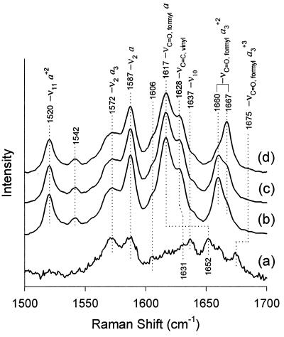
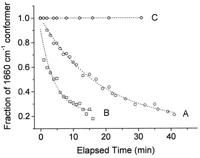
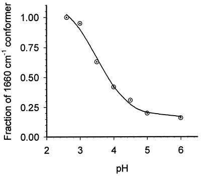
The hyperthermophilic archaeon Acidianus ambivalens expresses a membrane-bound aa(3)-type quinol oxidase, when grown aerobically, that we have studied by resonance Raman spectroscopy. The purified aa(3) oxidase, which does not contain bound quinol, undergoes a reversible slow conformational change at heme a(3) upon reduction, as indicated by a change in the frequency of its heme formyl stretching mode, from 1,660 cm(-1) to 1,667 cm(-1). In contrast, upon reduction of the integral membrane enzyme or the purified enzyme preincubated with decylubiquinol, this mode appears at 1,667 cm(-1) much more rapidly, suggesting a role of the bound quinol in controlling the redox-linked conformational changes. The shift of the formyl mode to higher frequency is attributed to a loss of hydrogen bonding that is associated with a group having a pKa of approximately 3.8. Based on these observations, a crucial element for proton translocation involving a redox-linked conformational change near the heme a(3) formyl group is postulated.


Figures

Figure 1
Figure 1
Resonance Raman spectra of reduced aa3 in the high-frequency region as a function of time. The excitation frequency was 413.1 nm, and the laser power at the sample was ≈10 mW. The purified enzyme (60 μM) was reduced anaerobically by dithionite, in 100 mM Mes, pH 6.0, 1 mg/ml lauryl maltoside. The spectra shown are (b) immediately (≈1 min) after reduction, (c) after 15 min, and (d) after 50 min. Also shown for comparison is the spectrum of the oxidized enzyme (spectrum a, laser power ≈0.1 mW).
Figure 2
Figure 2
Effect of quinol on the rate of conformational change in heme a3. The decrease in the heme a3 formyl line at 1,660 cm−1 was monitored as a function of time after reduction, in the presence and in the absence of quinol, at two different pH values. The enzyme was reduced anaerobically by dithionite at pH 6, 100 mM Mes, in the absence (A) and in the presence (B) of 1.6 mg/ml decylubiquinol. At pH 2.6 (C), the population of the 1,660-cm−1 conformer remained invariant in both the presence and the absence of decylubiquinol. The relative population of the 1,660-cm−1 (to that of 1,667 cm−1) conformer was calculated by curve fitting using two Lorentzians.
Figure 3
Figure 3
Variation in the population of the 1,660-cm−1 conformer as a function of pH (in the absence of decylubiquinol). The relative population of the 1,660-cm−1 conformer was calculated as in Fig. 2. Immediately after reduction, the 1,660-cm−1 conformer is the only species present at all pH values. The population of the 1,660-cm−1 conformer observed 100 min after reduction is shown (in ⊙). The sigmoidal curve (solid line) indicates the best fit that yielded an apparent pKa of 3.8 ± 0.4.
Figure 4
Figure 4
Structural model of the redox centers in A. ambivalens aa3 oxidase. The model was created based on the structural coordinates of Paracoccus cytochrome c oxidase, taking only the first 491 residues of the amino acid sequence into consideration. The residues are numbered as in bovine sequence. Shown in the picture are heme a, heme a3 (red), CuB (blue), and some of the neighboring residues (in thick lines), His-290 (one of the three histidine ligands to CuB), Tyr-244, Asp-364, His-368, Arg-438, and Arg-439. The residues shown in thin lines but not labeled are the histidine ligands to hemes a and a3 and CuB. The arrows denote the proposed path of proton translocation from His-290 to the Asp-364–propionate pair to the Arg-438–propionate region.
Figure 5
Figure 5
Model of proton pumping. The residues are numbered following the sequence of bovine aa3. The sequence of structures shown is I, the oxidized form; II, the transient reduced form; and III, the final reduced form. Only selective residues around the heme a3-CuB site are shown. Two unidentified groups capable of donating protons are represented as −BH and −AH. The red proton shown in bold denotes the translocable proton. Asp-364 and the propionate share the proton placed in between them. In the oxidized form (structure I), the formyl of a3 is not hydrogen-bonded as the H290 side chain is an imidazolate. Upon reduction, the formyl group forms hydrogen bonding transiently with the −NδH of imidazole (structure II). In structure III, the hydrogen bond is broken again and the proton is transferred to the propionate–Asp-364 pair.

Similar articles

Cited by

References

    1. Anemuller S, Schmidt C L, Pacheco I, Schafer G, Teixeira M. FEMS Microbiol Lett. 1994;117:275–280.
    1. Schafer G. Biochim Biophys Acta. 1996;1277:163–200. - PubMed
    1. Zillig W, Yeats S, Holz I, Bock A, Gropp F, Rettenberger M, Lutz S. Nature (London) 1985;313:789–791. - PubMed
    1. Fuchs T, Huber H, Burggraf S, Stetter K O. Syst Appl Microbiol. 1996;19:56–60.
    1. Giuffre A, Gomes C M, Antonini G, D’Itri E, Teixeira M, Brunori M. Eur J Biochem. 1997;250:383–388. - PubMed

Publication types

LinkOut - more resources