

doi: 10.1073/pnas.96.17.9628.
Basonuclin, a zinc finger protein of keratinocytes and reproductive germ cells, binds to the rRNA gene promoter
Affiliations
- PMID: 10449744
- PMCID: PMC22260
- DOI: 10.1073/pnas.96.17.9628
Item in Clipboard
Basonuclin, a zinc finger protein of keratinocytes and reproductive germ cells, binds to the rRNA gene promoter
Proc Natl Acad Sci U S A.
.
Abstract
Basonuclin is a protein containing three pairs of C(2)H(2) zinc fingers. The protein has been found in the basal (germinal) cell layer of stratified squamous epithelia, such as the epidermis, and in germ cells of the testis and ovary. We show here that the human protein has specific affinity for a segment of the promoter of the gene for rRNA. Basonuclin interacts with two separate parts of the promoter, each possessing dyad symmetry. The upstream part, but not the downstream part, is known to bind UBF1, a transcription factor for rDNA. Basonuclin is likely to be a cell-type-specific regulatory protein for rDNA transcription.
Figures
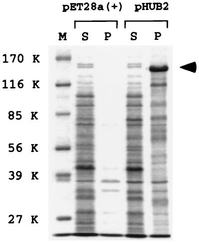
Recombinant basonuclin recovered from inclusion bodies. E. coli expressing human basonuclin from plasmid pHUB2 were disrupted, and the lysate was divided into soluble and particulate fractions (S and P). Each fraction was subjected to SDS/PAGE, and the resolved proteins were stained with Coomassie blue. Human basonuclin, indicated by an arrowhead at 140 kDa (K), was the most abundant protein. This protein was absent from the particulate fraction of cells harboring the vector plasmid pET28a(+). Basonuclin was absent from both soluble fractions, which otherwise contained virtually identical proteins. M, standard markers.
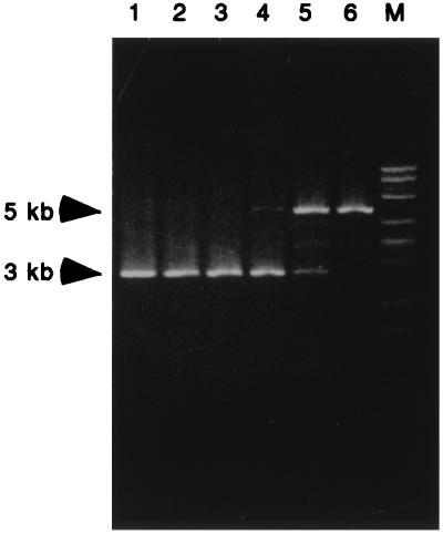
A human genomic fragment with affinity for basonuclin. A genomic library was subjected to selection based on affinity binding to basonuclin inclusion bodies. After each cycle of binding and elution, the DNA was amplified, digested with KpnI, and analyzed by agarose electrophoresis and ethidium bromide staining. The number above each lane indicates the selection cycle. After the fifth cycle, a single band of 5 kilobases (kb) became dominant (pTRG1). The band of 3 kb, corresponding to the size of the vector, could no longer be detected after the sixth cycle. In lane M, a λDNA–BstEII digest is shown as standard marker.
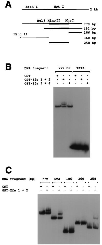
Binding of the pTRG1 insert and its subfragments to Zfs of basonuclin. (A) Schematic of the subcloned human genomic DNA. The left end of the 360-bp fragment was created by digesting the vector sequence with HincII instead of EcoRI. (B) Gel-shift assay of binding between the 779-bp DNA fragment and Zfs coupled to GST. GST–Zfs 1+2 (40 ng), GST–Zfs 3+4 (60 ng), and GST itself (40 ng) were each incubated with the 32P-labeled 779-bp fragment (7.5 fmol), and the mobility of the DNA was tested by electrophoresis through a 4% nondenaturing polyacrylamide gel. A 32P-labeled 25-nucleotide oligomer containing TATA (17 fmol) served as a control. GST–Zfs 1+2 bound to the 779-bp fragment, but GST–Zfs 3+4 did not. No Zfs bound to the fragment containing TATA. (C) GST–Zfs 1+2 binding to subfragments. GST–Zfs 1+2 (40 ng) or GST itself (40 ng) was incubated with each labeled fragment (4,500 cpm; 779-, 492-, 186-, 360-, or 258-bp DNA), and the mobility of the DNA was assayed. A binding site for GST–Zfs 1+2 was present in the 799-, 492-, and 258-bp fragments, as indicated by thick lines in A, but not in the 186- or 360-bp fragments.
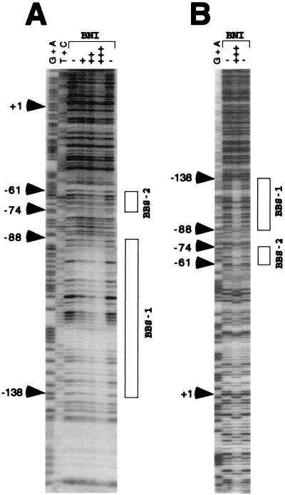
Resistance to DNase I of rRNA promoter region bound to basonuclin inclusion bodies (BNI). (A) The noncoding strand (24 fmol) of the 258-bp DNA of the human rRNA promoter, to which a short stretch of vector sequence was added to label its 3′ end, was incubated with (+, 5 μg; ++, 10 μg; +++, 20 μg) or without (−) BNI and then digested with 10 milliunits of DNase I. BNI protected the BBS1 region and, less strongly, BBS2 (shown by boxes). The G + A and T + C sequence reactions were performed as described (11). (B) The coding strand (43 fmol) of the EcoRI–NheI fragment of the human rRNA promoter, labeled at the 3′ end, was incubated with (+++, 20 μg) or without (−) BNI and then digested with 12.5 milliunits of DNase I. Both BBS1 and BBS2 were protected by BNI. BBS2 was more strongly protected in this strand than in the noncoding strand.
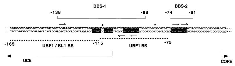
Location of binding sites of basonuclin relative to those of UBF1. The sequence shown is that of the promoter region of the human rRNA gene (18). The upper strand is the RNA-like (coding) strand. The CORE region is essential for transcription but is not protected from nuclease digestion by either UBF1 or basonuclin. BBS1 and BBS2 are indicated by boxes. BBS1 contains a dyad sequence (black background), whose center is indicated by a black dot. BBS2 contains a dyad sequence whose complement is located near the 3′ end of BBS1. The axis of symmetry, indicated by a triangle, is located between nucleotides −82 and −83. The intervening 18-bp spacer consisting of 15 nucleotides is not protected from DNase I. The UBF1-binding region is shown by asterisks below the sequence. The 3′ half is protected by UBF1 alone (upper row of asterisks), and the 5′ half is protected only when SL1 is present as well (lower row of asterisks). BBS1 is centered almost at the same point as the entire binding region of UBF1. BBS2 is not protected by UBF1. Arrows indicate the repeated sequence GCGA, which occurs in both strands of the dyad sequence.
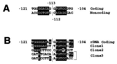
Homology between basonuclin-binding sites of human rRNA promoter and other genes. (A) BBS1. The dyad sequence, shown against a black background, with its axis of symmetry between positions −113 and −112. (B) The same sequence compared with homologous sequences of the three other selected DNA sequences (clones 1–3). Nucleotide identities are shown against a black background. In the case of clone 3, two sequences homologous to the sequence of rDNA were present.
References
Publication types
MeSH terms
Substances
LinkOut - more resources
Full Text Sources
Other Literature Sources
Molecular Biology Databases
