. 1999 Aug 17;96(17):9637-41.
doi: 10.1073/pnas.96.17.9637.

Immortalization of primary human keratinocytes by the helix-loop-helix protein, Id-1

Affiliations

Immortalization of primary human keratinocytes by the helix-loop-helix protein, Id-1

R M Alani et al. Proc Natl Acad Sci U S A. .

Abstract

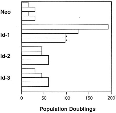
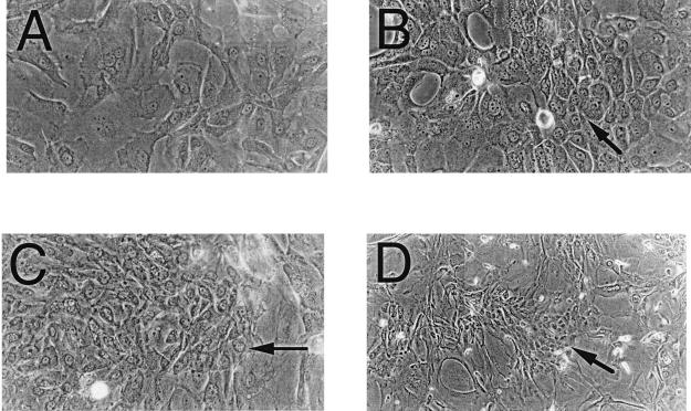
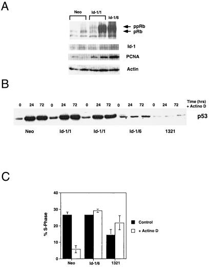
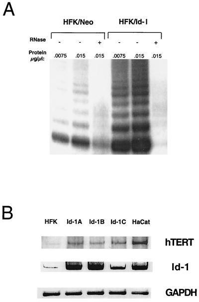
Basic helix-loop-helix (bHLH) DNA-binding proteins have been demonstrated to regulate tissue-specific transcription within multiple cell lineages. The Id family of helix-loop-helix proteins does not possess a basic DNA-binding domain and functions as a negative regulator of bHLH proteins. Overexpression of Id proteins within a variety of cell types has been shown to inhibit their ability to differentiate under appropriate conditions. We demonstrate that ectopic expression of Id-1 leads to activation of telomerase activity and immortalization of primary human keratinocytes. These immortalized cells have a decreased capacity to differentiate as well as activate phosphorylation of the retinoblastoma protein. Additionally, these cells acquire an impaired p53-mediated DNA-damage response as a late event in immortalization. We conclude that bHLH proteins play a pivotal role in regulating normal keratinocyte growth and differentiation, which can be disrupted by the immortalizing functions of Id-1 through activation of telomerase activity and inactivation of the retinoblastoma protein.


Figures

Figure 1
Figure 1
Population doublings of Id-transfected primary human keratinocytes. Population doublings were determined for four separate transfections. All cells were passaged continuously since the day of transfection (after 20–30 doublings). Two of the Id-1 cell lines were contaminated after 6 months of passage (∗). Two additional cell lines were frozen down after 8 months and 1 year of passage.
Figure 2
Figure 2
Photomicrographs of Id-transfected HFKs under differentiating stimuli. Phase-contrast photomicrographs demonstrate focal regions of smaller, phenotypically less differentiated cells (arrows) in Id-1- (B), Id-2- (C), and Id-3- (D) transfected HFKs vs. vector control-transfected cells (A), which show a typical differentiated phenotype of large, flattened cells with increased cytoplasmic-to-nuclear ratios.
Figure 3
Figure 3
(A) Immunoblot of pRb, proliferating cell nuclear antigen, and Id-1 expression in Id-1 transfectants. Neo, vector-transfected HFKs 1 month after transfection; Id-1/1, Id-1-transfected HFKs 1 month after transfection; Id-1/6, Id-1-transfected HFKs 6 months after transfection. Actin panel is used as a loading control. (B) Immunoblot of p53 expression in response to DNA damage by actinomycin D for 24 and 72 hr. Neo cells are vector-transfected HFKs 1 month after transfection, Id-1/1 cells are Id-1-transfected HFKs 1 month after transfection, Id-1/6 cells are Id-1-transfected HFKs 6 months after transfection, and 1321 cells are HPV 16 E6/E7-immortalized HFKs. (C) Fluorescence-activated cell sorter analysis of Id-1-immortalized HFK response to DNA damage. Cells were exposed to 2.5 nM actinomycin D for 48 hr, and cell cycle profiles were determined by propidium iodide staining. Neo cells are vector-transfected HFKs, Id-1/6 are Id-1-transfected HFKs 6 months after transfection, and 1321 cells are HPV 16 E6/E7 immortalized HFKs.
Figure 4
Figure 4
(A) Telomerase activity in Id-1-expressing human keratinocytes. Normal human keratinocytes were transfected with either vector alone (HFK-Neo) or with a vector expressing Id-1 (HFK-Id). Cells were selected for expression of neomycin resistance and were analyzed for telomerase activity by the TRAP assay at 1 month after transfection. (B) hTERT expression in Id-1-transfected human keratinocytes. hTERT and Id-1expression was evaluated in normal human keratinocytes (HFK) and Id-1-transfected keratinocytes at 1 month (Id-1A, Id-1B) and 1 year (Id-1C) after transfection by using RT-PCR analysis. The spontaneously immortalized human keratinocyte cell lines, HaCaT, were used as a positive control for hTERT expression. Glyceraldehyde-3-phosphate dehydrogenase expression is shown as an internal control.

References

    1. Olson E N, Klein W H. Genes Dev. 1994;8:1–8. - PubMed
    1. Weintraub H. Cell. 1993;75:1241–1244. - PubMed
    1. Benezra R, Davis R L, Lockshon D, Turner D L, Weintraub H. Cell. 1990;61:49–59. - PubMed
    1. Sun X H, Copeland N A, Jenkins N A, Baltimore D. Mol Cell Biol. 1991;11:5603–5611. - PMC - PubMed
    1. Wilson R B, Kiledjian M, Shen C P, Benezra R, Zwollo P, Dymecki S M, Desiderio S V, Kadesch T. Mol Cell Biol. 1991;11:6185–6191. - PMC - PubMed

Publication types