. 1999 Aug 17;96(17):9648-53.
doi: 10.1073/pnas.96.17.9648.

Wiskott-Aldrich syndrome protein regulates podosomes in primary human macrophages

Affiliations

Wiskott-Aldrich syndrome protein regulates podosomes in primary human macrophages

S Linder et al. Proc Natl Acad Sci U S A. .

Abstract

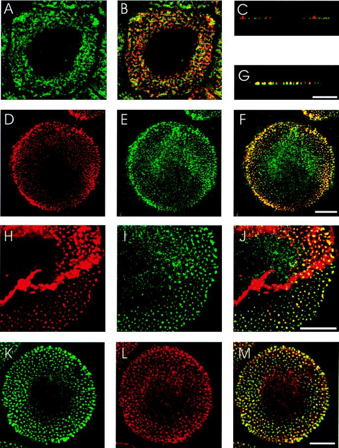
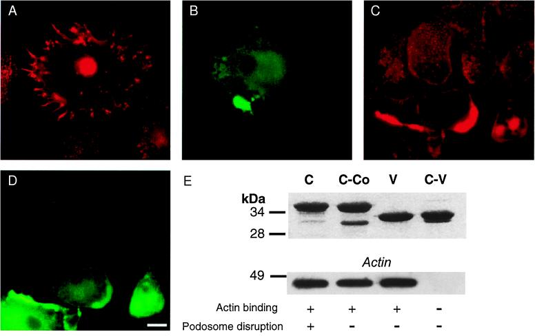
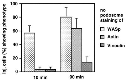
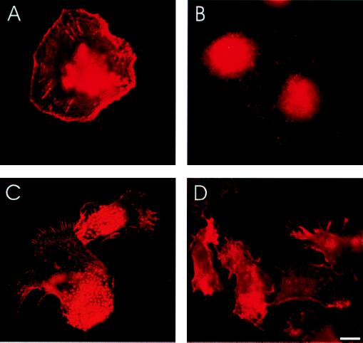
Wiskott-Aldrich syndrome protein (WASp) is a hematopoietic-specific, multidomain protein whose mutation is responsible for the immunodeficiency disorder Wiskott-Aldrich syndrome. WASp contains a binding motif for the Rho GTPase CDC42Hs as well as verprolin/cofilin-like actin-regulatory domains, but no specific actin structure regulated by CDC42Hs-WASp has been identified. We found that WASp colocalizes with CDC42Hs and actin in the core of podosomes, a highly dynamic adhesion structure of human blood-derived macrophages. Microinjection of constitutively active V12CDC42Hs or a constitutively active WASp fragment consisting of the verprolin/cofilin-like domains led to the disassemly of podosomes. Conversely, macrophages from patients expressing truncated forms of WASp completely lacked podosomes. These findings indicate that WASp controls podosome assembly and, in cooperation with CDC42Hs, podosome disassembly in primary human macrophages.


Figures

Figure 1
Figure 1
Localization of actin, vinculin, CDC42Hs, and WASp in podosomes of primary human macrophages. Confocal laser scanning micrographs showing horizontal sections of ventral parts of cells (A, B, D–F, and H–M), vertical sections of peripheral parts of cells (C and G), vinculin staining(A), overlays of vinculin (green) and actin staining (red) (B and C), actin staining (D), CDC42Hs staining (E), overlay of D and E (F), overlay of actin (red) and CDC42HS staining (G), actin staining (H), WASp staining (I) overlay of H and I (J), WASp staining (K), CDC42Hs staining (L), and overlay of K and L (M). Yellow color indicates colocalization of red and green. [Bar = 10 μm for each row.]
Figure 2
Figure 2
CDC42Hs and WASp control podosomes. Immunofluorescence micrographs of primary human macrophages. (A) Cell injected with V12CDC42Hs, actin staining; (B) injection control of A, staining against coinjected rat IgG. (C) Macrophages injected with C-terminal WASp polypeptide C, actin staining; (D) injection control of C, staining against coinjected rat IgG. [Bar = 10 μm.] (E) (Upper) GST-fusions of C-terminal WASp polypeptides. SDS/polyacrylamide gel stained with Coomassie blue. Denominations of polypeptides are given above each lane. Running behavior of polypeptides differs in some cases from theoretical molecular mass, which is indicated on the left in kDa. (Lower) Actin binding of C-terminal WASp polypeptides. Western blot probed with actin-specific antibody. Molecular mass is indicated on the left in kDa. Abilities of polypeptides to bind actin and disrupt podosomes are indicated below each lane by + or −.
Figure 3
Figure 3
Time course of podosome disruption after injection of C-terminal WASp domain. Cells were fixed at the indicated time points after injection. Percentage of cells without podosomal staining of the respective protein is represented by bars. For each value, three separate injections of 30 cells were performed. Standard errors are indicated by error bars. Uninjected cells or cells injected with GST show values below 3% for loss of podosomal staining for each protein (data not shown).
Figure 4
Figure 4
WAS macrophages have defects in podosome formation and cell polarization. Immunofluorescence micrographs of primary human macrophages. (A) Macrophage of WAS patient 3603, actin staining. (B) Macrophages of WAS patient 3,603, vinculin staining. (C and D) Macrophages stimulated with fMLP, actin staining. (C) Macrophages from healthy donor. (D) Macrophages from WAS patient 3,603. [Bar = 10 μm.]

References

    1. Kirchhausen T. Mol Med Today. 1998;9:300–304. - PubMed
    1. Kenney D M, Cairns L, Remold-O′Donnel E, Peterson J, Rosen F S, Parman R. Blood. 1986;68:1329–1332. - PubMed
    1. Remold-O’Donnell E, Rosen F S, Kenney D M. Blood. 1996;87:2621–2631. - PubMed
    1. Ochs H D. Springer Semin Immunopathol. 1998;19:459–478. - PubMed
    1. Binks M, Jones G E, Brickell P M, Kinnon C, Katz D R, Thrasher A J. Eur J Immunol. 1998;28:3259–3267. - PubMed

Publication types

MeSH terms

LinkOut - more resources