. 1999 Aug 17;96(17):9751-6.
doi: 10.1073/pnas.96.17.9751.

Two major forms of DNA (cytosine-5) methyltransferase in human somatic tissues

Affiliations

Two major forms of DNA (cytosine-5) methyltransferase in human somatic tissues

D W Hsu et al. Proc Natl Acad Sci U S A. .

Abstract

Thus far, only one major form of vertebrate DNA (cytosine-5) methyltransferase (CpG MTase, EC 2.1.1.37) has been identified, cloned, and extensively studied. This enzyme, dnmt1, has been hypothesized to be responsible for most of the maintenance as well as the de novo methylation activities occurring in the somatic cells of vertebrates. We now report the discovery of another abundant species of CpG MTase in various types of human cell lines and somatic tissues. Interestingly, the mRNA encoding this CpG MTase results from alternative splicing of the primary transcript from the Dnmt1 gene, which incorporates in-frame an additional 48 nt between exons 4 and 5. Furthermore, this 48-nt exon sequence is derived from the first, or the most upstream, copy of a set of seven different Alu repeats located in intron 4. The ratios of expression of this mRNA to the expression of the previously known, shorter Dnmt1 mRNA species, as estimated by semiquantitative reverse transcription-PCR analysis, range from two-thirds to three-sevenths. This alternative splicing scheme of the Dnmt1 transcript seems to be conserved in the higher primates. We suggest that the originally described and the recently discovered forms of CpG MTase be named dnmt1-a and dnmt1-b, respectively. The evolutionary and biological implications of this finding are discussed in relation to the cellular functions of the CpG residues and the CpG MTases.


Figures

Figure 1
Figure 1
Structural map of human dnmt1. The structure of the human CpG MTase dnmt1 is shown schematically at the cDNA level. The numbering of nucleotide sequences (1–5,387) follows that used in ref. . Besides the initiation and termination codons, the locations of the cDNA regions coding for the PCNA-binding domain, the nuclear localization signal, and the replication foci-targeting sequence are also indicated. The site of insertion of the 48-bp Alu repeat segment through alternative splicing is also marked with a black triangle. It is between nucleotides 682 and 683. UTR, untranslated region.
Figure 2
Figure 2
Identification of alternative splicing of human Dnmt1 transcript. (A) A 2% agarose gel electrophoresis of RT-PCR products of human K562 RNA. RT-PCR was carried out as described in Materials and Methods by using the PCR primer pair 502–521 and 1,001–981. Lane M, DNA length marker; lane 1, PCR product without RT reaction; lane 2, RT-PCR product. (B) Nucleotide sequences of the two bands, 550 bp and 500 bp, from lane 2 in A. The sequence of the coding strand of the 500-bp fragment from position 607 to 756 is shown together with the corresponding amino acids. Sequence of the inserted 48-bp segment in the 550-bp fragment is aligned with the homologous region of the antisense strand of the Alu consensus sequence described in ref. . Note that the 48-bp insertion results in the substitution of proline (P) at codon 149 by arginine (R), as well as the in-frame insertion of another 16 amino acids, serine (S) through alanine (A).
Figure 3
Figure 3
(A) A 2% agarose gel electrophoresis of RT-PCR products of human K562 RNA with the primer pair 238–254 and 5,130–5,101. (B) The incorporation of the 48-nt segment into a Dnmt1 transcript of an approximate length of 5,000 nt. Lane 1, PCR of the combined eluate from gel regions immediately above and below the Dnmt1 band in lane K562 in A; lane 2, PCR of the Dnmt1 band from lane K562 in A; lane 3, the same RT-PCR sample as in lane 2 of Fig. 2A. (C) RT-PCR “scanning” of Dnmt1 transcript. PCRs were carried out with different pairs of primers. The numbers above each lane indicate the nucleotide positions of the ends of DNA fragments amplified. Note that the alternative splicing of 48 nt is confirmed further by the appearance of double bands in both the second lane (nucleotides 238–718) and the third lane (nucleotides 601–1,001).
Figure 4
Figure 4
Relative expression of Dnmt1-a and Dnmt1-b in total RNAs from K562, HeLa, and NK cells. The analysis was performed as described in detail in Materials and Methods. HD indicates the band resulting from heteroduplex formation between Dnmt1-b and Dnmt1-a during the PCR cycles (data not shown). The RT-PCR samples loaded were derived from 10 ng (lanes 1), 5 ng (lanes 2), and 2.5 ng (lanes 3) of total RNA.
Figure 5
Figure 5
Enrichment of both Dnmt1-a and Dnmt1-b in poly(A)-RNA. RT-PCR product from 0.25 ng of K562 poly(A)-RNA (lane 2) was loaded, along with, for purposes of comparison, that from 2 ng of K562 total RNA (lane 1).
Figure 6
Figure 6
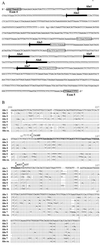
(A) Nucleotide sequence of intron 4 of human Dnmt1 gene. Only the coding strand is shown; exon 4, exon 5, and locations of different Alu family repeats within the intron are indicated. The sequence of approximately 300 bp (in parentheses) between Alu2 and Alu3 could not be determined accurately. The 7-bp sequence in Alu4 is also uncertain. The direct repeats flanking Alu3 and the tetrameric array composed of Alu4, Alu5, Alu6, and Alu7 are in individual boxes. (B) Alignment of the seven Alu family repeats of human Dnmt1 intron 4 and the Alu consensus sequence (Alu co.). Only sequences of the antisense strands are shown. The numbering follows that used for the Alu consensus in ref. . The segment of Alu1 is shown in full. Nucleotides identical to Alu1 are represented by the vertical bar. Relative deletions are indicated by hyphens. Note that Alu5 is only half the length of a typical monomeric Alu repeat and could have resulted from deletion via homologous recombination within an ancestral Alu sequence. The 48 nt in Alu1 that could be alternatively spliced to generate Dnmt1-b RNA are indicated by bold letters. Also shown above the junction between this 48-nt segment and its flanking regions are the consensus sequences of the acceptor and donor sites for eukaryotic mRNA splicing.
Figure 7
Figure 7
RT-PCR comparison of total RNAs isolated from human K562 and chimpanzee blood. The primers 502–521 and 1,001–981 were used for PCR, and the products were analyzed on a 2% agarose gel.
Figure 8
Figure 8
Summary of the alternative splicing schemes of human Dnmt1 transcript for the generation of Dnmt1-a (B Lower) and Dnmt1-b (B Upper). The genomic map of the Dnmt1 gene is shown in A with exons 1–11 and 40 indicated.

Similar articles

Cited by

References

    1. Jost J P, Saluz H P, editors. DNA Methylation: Molecular Biology and Biological Significance. Basel: Birkhauser; 1993.
    1. Bestor T H, Verdine G L. Curr Opin Cell Biol. 1994;6:386–389. - PubMed
    1. Bestor T H. Nature (London) 1998;393:311–312. - PubMed
    1. Jones P A, Buckley J D. Adv Cancer Res. 1990;54:1–23. - PubMed
    1. Baylin S B. Science. 1997;277:1948–1949. - PubMed

Publication types

Associated data