. 1999 Aug 17;96(17):9885-90.
doi: 10.1073/pnas.96.17.9885.

Mammalian ELAV-like neuronal RNA-binding proteins HuB and HuC promote neuronal development in both the central and the peripheral nervous systems

Affiliations

Mammalian ELAV-like neuronal RNA-binding proteins HuB and HuC promote neuronal development in both the central and the peripheral nervous systems

W Akamatsu et al. Proc Natl Acad Sci U S A. .

Abstract

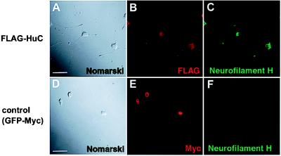
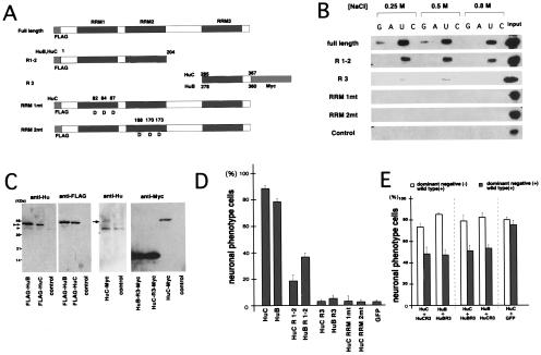
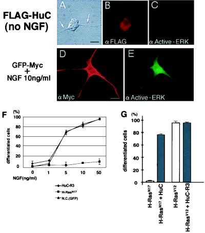
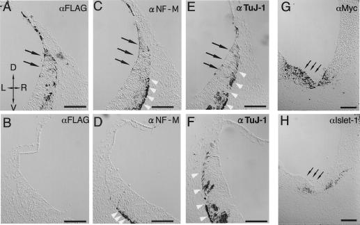
Hu proteins are mammalian embryonic lethal abnormal visual system (ELAV)-like neuronal RNA-binding proteins that contain three RNA recognition motifs. Although Drosophila ELAV is required for the correct differentiation and survival of neurons, the roles played by the Hu genes in the mammalian nervous system remain largely unknown. To explore the in vivo functions of mouse Hu proteins, we overexpressed them in rat pheochromocytoma PC12 cells, where they induced neuronal phenotype in the absence of nerve growth factor. We have characterized the functions of various forms of mHuB and mHuC bearing point mutations or deletions. Mutants of mHuC that had amino acid exchanges in the RNP1 domain of the first or second RNA recognition motifs (RRMs) lost biologic activity as well as RNA-binding activity. In addition, the mutants containing only the third RRM failed to induce the neuronal phenotype in PC12 cells and inhibited the biologic activity of cotransfected wild-type mHuB and mHuC, thus acting as a dominant-negative form. However, these mutants could not suppress the nerve growth factor-induced differentiation of PC12 cells. Further, we misexpressed wild-type and dominant-negative Hu in E9.5 mouse embryos, by using electroporation into the neural tube at the level of the rhombencephalon. mHuB and mHuC induced the ectopic expression of neuronal markers, whereas the dominant-negative forms of mHuB and mHuC suppressed the differentiation of central nervous system motor neurons. From these results, we suggest that Hu proteins are required for neuronal differentiation in the mammalian nervous system.


Figures

Figure 1
Figure 1
HuC-induced neuronal phenotype of PC12 cells. (A) Morphology of pCXN2-FLAG-HuC-transfected PC12 cells (Nomarski differential interference contrast optics). Neuron-like morphology, similar to NGF-induced differentiation, appeared 9 d after transfection. (D–F) Negative control (pCXN2-GFP-Myc transfected PC12 cells) showed no morphological changes 9 d after transfection. Double staining for overexpressed FLAG-HuC (or GFP-Myc) (B and E) and neurofilament H (C and F). Antibodies for tagged sequence were used for the detection of transfected fusion protein. Overexpressed HuC protein localized mainly to the cytoplasm, and these cells showed increased expression of Neurofilament H (C), which is also known to increase in the NGF-induced differentiation of PC12 cells (53). (Bar = 15 μm.)
Figure 2
Figure 2
Mutation analyses of Hu (HuB and HuC) proteins. (A) Mutants of Hu proteins used for functional analysis. HuB and C deletion mutants R1–2 and R3, and HuC RRM amino acid replaced mutants (RRM1mt, RRM2mt) were constructed as described in Material and Methods. (B) RNA-binding assay of HuC mutants. Full-length and mutant HuC fusion proteins and a control fusion protein were incubated with poly(rG)-, poly(rA)-, poly(rU)-, and poly(rC)-Sepharose RHP beads in either 0.25 M, 0.5 M, or 0.8 M NaCl. After being washed, protein bound to the RHP was analyzed by Western blot analysis with an anti-T7 antibody (Novagen). (C) Expression of exogenous and endogenous Hu proteins in PC12 cells 2 d after transfection. To detect these proteins, immunoblot analyses were performed. The expression level of exogenous FLAG-tagged full- length transgene products (indicated by double arrowheads) were quantitated in comparison to the endogenous Hu proteins of control cells (pCXN2 transfected PC12 cells) (indicated by arrowhead). The expression level of Myc-tagged HuB/C-R3 transgene products was indirectly quantitated by comparing to those of Myc-tagged full length transgene product (indicated by small arrow) and endogenous Hu proteins in the control cells. (D) Neuronal differentiation-inducing activities of PC12 cells by Hu mutants. The differentiation-inducing activities of mutants were evaluated by observing the morphology of transfected PC12 cells. G418 was added to the medium to enrich Hu-transfected cells. The cells that had dendrites longer than the diameter of their cell bodies were termed “neuronal phenotype” cells. This graph shows the proportion of differentiated cells among the transgene-containing cells (detected by using antibodies against the FLAG or Myc tags) 9 d after transfection. FLAG-GFP fusion protein was used as a negative control. One hundred transgene-expressing cells were examined three times (total 300 cells) per construct. (E) Hu-R3 mutants act as dominant-negative forms of Hu. Full-length Hu and Hu-R3 mutants were cotransfected into PC 12 cells and analyzed by using the same criteria as in D (single positive and double positive, each 100 cells, three times). Differentiation of cells transfected with both full-length Hu and the Hu-R3 mutant (double-positive cells) was reduced compared with cells transfected with full-length Hu alone (single-positive cells). Transfection of HuC with HuB-R3 or HuB with HuC-R3 showed the same results. Negative controls (pCXN2-FLAG-HuC with pCXN2-GFP-Myc) did not show any inhibition of neuronal differentiation.
Figure 3
Figure 3
Hu proteins act independently of the NGF/Ras cascade. (A–E) HuC-induced neuronal phenotype of PC12 cells is not accompanied by ERK activation. Expression of activated-ERK in PC12 cells overexpressing HuC in the absence of additional NGF (A–C) or cells expressing GFP-Myc in the presence of additional NGF (D and E) 9 d after transfection. There was no detectable expression of activated ERK in neuron-like PC12 cells that overexpressed HuC. Bar = 5 μm. (F) HuC-R3 cannot prevent NGF-induced PC12 differentiation. PC12 cells transfected with HuC-R3, H-RasN17 (dominant negative) or FLAG-GFP (negative control) were cultured in medium containing additional NGF. Transfected cells were selected by using G418. Morphology of transfected cells was evaluated 9 d after transfection same as in Fig. 3. Although H-RasN17 misexpression almost completely blocked NGF-induced differentiation, HuC-R3 misexpression showed no significant influence on differentiation. (G) Dominant-negative H-Ras cannot prevent HuC-induced neuronal phenotype. H-RasN17 and HuC, or H-RasV12(dominant active) and HuC-R3 were cotransfected into PC12 cells. Morphology of the cells expressing both proteins was evaluated as in the legend for Fig. 2. Dominant-negative H-Ras (H-RasN17) could not suppress HuC-induced neuronal phenotype, and HuC-R3 could not suppress dominant active Ras (H-RasV12)-induced differentiation.
Figure 4
Figure 4
Misexpression of Hu and Hu-R3 mutants in mouse embryonic CNS. Twelve-micrometer frozen serial sections of a HuC (FLAG-tagged)-transfected E9.5 mouse embryo (A–F) and a HuC-R3 (Myc-tagged)-transfected embryo (G and H) (D, dorsal; V, ventral; L, left; R, right). Sections of the HuC-introduced embryo were immunostained with antibodies against FLAG (A and B), neurofilament M (C and D) and TuJ-1 (E and F). Paired images of each individual section are shown from the transfected (A, C, and E) and nontransfected (B, D, and F) sides of the same slices. At the rhombencephalon of the normal mouse embryos in these stages (E9.5), the neurogenesis has already begun and neuronal markers are seen. Therefore, even in the nontransfected side (D and F), the expressions of authentic neuronal markers [TuJ1 (D); NF-M (F)] were observed in the outside of the ventricular zone (indicated by white arrowheads), corresponding to normal neuronal development. In the transfected side (C and E), however, ectopically induced expressions of neuronal markers were observed from within the ventricular zone [indicated by black arrows, TuJ-1 (C); NF-M (E)], coinciding with the expression of transfected FLAG-HuC [indicated by black arrows in (A)]. Such ectopic expressions of neuronal markers are never seen in normal neuronal development. Sections of a HuC-R3-transfected embryo were immunostained by using antibodies against Myc-tag (G) and Islet-1 (H). Islet-1-positive cells decreased in the HuC-R3 overexpressing region (indicated by small arrows). (Bar = 100 μm.)

References

    1. Okano H. Dev Growth Differ. 1995;37:619–629. - PubMed
    1. Liu Q, Siomi H, Siomi M C, Fischer U, Zhang Y, Wan L, Dreyfuss G. Cold Spring Harbor Symp Quant Biol. 1996;61:689–697. - PubMed
    1. Sakakibara S, Okano H. J Neurosci. 1997;17:8300–8312. - PMC - PubMed
    1. Grabowski P J. Cell. 1998;92:709–712. - PubMed
    1. Robinow S, Campos A R, Yao K M, White K. Science. 1988;242:1570–1572. - PubMed

Publication types

MeSH terms

LinkOut - more resources