. 1999 Aug 17;96(17):9891-6.
doi: 10.1073/pnas.96.17.9891.

theta, a novel gamma-aminobutyric acid type A receptor subunit

Affiliations

theta, a novel gamma-aminobutyric acid type A receptor subunit

T P Bonnert et al. Proc Natl Acad Sci U S A. .

Abstract

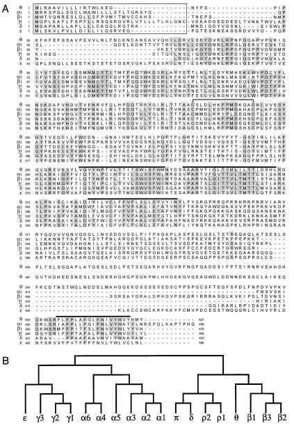
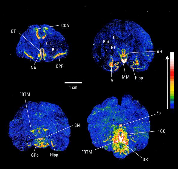
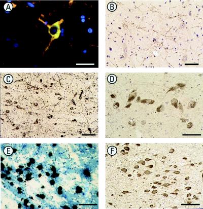
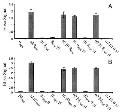
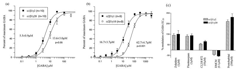
gamma-Aminobutyric acid type A (GABA-A) receptors are a major mediator of inhibitory neurotransmission in the mammalian central nervous system, and the site of action of a number of clinically important drugs. These receptors exist as a family of subtypes with distinct temporal and spatial patterns of expression and distinct properties that presumably underlie a precise role for each subtype. The newest member of this gene family is the theta subunit. The deduced polypeptide sequence is 627 amino acids long and has highest sequence identity (50.5%) with the beta1 subunit. Within the rat striatum, this subunit coassembles with alpha2, beta1, and gamma1, suggesting that gamma-aminobutyric acid type A receptors consisting of arrangements other than alpha beta + gamma, delta, or epsilon do exist. Expression of alpha2beta1gamma1theta in transfected mammalian cells leads to the formation of receptors with a 4-fold decrease in the affinity for gamma-aminobutyric acid compared with alpha2beta1gamma1. This subunit has a unique distribution, with studies so far suggesting significant expression within monoaminergic neurons of both human and monkey brain.


Figures

Figure 1
Figure 1
Human GABA-A receptor θ subunit. (A) Alignment of the deduced amino acid sequences of the human GABA-A receptor α1(18), β1(18), γ1 (19), δ (unpublished results), ɛ (4), and θ subunits. Numbering of amino acids is given by assigning the initiating methionine as 1. Positions where amino acid residues are conserved in four or more sequences are shaded. Putative signal peptides (20) are boxed, putative transmembrane domains are overlined, and the two cysteine residues conserved in the ligand-gated ion channel family are joined by a solid line. (B) Dendrogram of the deduced amino acid sequences of the GABA-A receptor (and ρ1–ρ2 of GABA-C) family. The analysis was performed with clustalw (GCG).
Figure 2
Figure 2
In situ hybridization autoradiograms of monkey brain sections showing expression of θ subunit mRNA. The signal intensity is indicated, with white representing the strongest signal. CCA, anterior cingulate cortex; OT, olfactory tract; NA, nucleus accumbens; Cd, caudate; Put, putamen; CPF, piriform cortex; GP, globus pallidus; AH, anterior hypothalamus; Hipp, hippocampus; MM, mamillary body; A, amygdala; FRTM, reticular formation; GPo, pons; DR, dorsal raphe nucleus; GC, central grey; Ep, pineal gland.
Figure 3
Figure 3
Immunohistochemical localization of the GABA-A receptor θ subunit in monkey and human brain. (A) The expression of immunoreactivity (green) for the GABA-A receptor θ subunit in the dopaminergic neurons of the periventricular hypothalamus of monkey brain was confirmed by colocalization with TH immunoreactivity (red). Colocalization is observed as yellow/orange. Nuclei are counterstained with Hoechst 33258 (blue). (Bar = 30 μm.) (B) θ subunit immunoreactive processes in the monkey globus pallidus are similar in pattern to those demonstrated by TH, further suggesting expression by nigra–striatal projection neurons. (Bar = 50 μm.) (C) θ subunit immunoreactivity is expressed by the neurons of the primate substantia nigra pars compacta, suggesting expression in dopaminergic neurons. (Bar = 10 μm.) (D) θ subunit immunoreactivity in melanized neurons of the human substantia nigra pars compacta, again suggesting expression in dopaminergic neurons. (Bar = 50 μm.) (E) High-resolution autoradiography in situ hybridization experiments show expression of θ subunit mRNA as silver grains over melanized neurons of the human locus coeruleus. (Bar = 100 μm.) (F) θ subunit immunoreactivity in human locus coeruleus melanized neurons (probably noradrenergic). (Bar = 100 μm.)
Figure 4
Figure 4
Coassembly and cell surface expression of θ with other GABA-A receptor subunits. c-myc epitope tagged θ (A) or c-myc-epitope-tagged β2 (B) was transiently transfected into HEK 293 cells, either alone or with other GABA-A receptor subunits. Cell surface expression of θ-myc or β2-myc was measured by a whole cell surface ELISA assay. The ELISA signal (A620) is proportional to the amount of θ-myc or β2-myc at the cell surface. Background signal from mock-transfected control wells has been subtracted, and values are the mean ± SEM of triplicate determinations.
Figure 5
Figure 5
Functional effects of θ expressed with other GABA-A receptor subunits. α1β1γ2 (a) and α2β1γ1 (b) GABA-A subunit cDNAs were transiently transfected both with (○) and without (■) the θ subunit cDNA into HEK 293 cells. Whole-cell patch-clamp techniques were used to measure currents in response to GABA after receptor expression. Concentration–response curves to GABA are shown normalized to a maximum response. Data are the mean ± SEM. of the number of cells indicated. Values are the mean EC50 ± SEM calculated from fits to individual cells. (c) Modulation of the GABA EC20 response of α1β1γ2 and α1β1γ2θ expressed in oocytes by a maximum concentration of benzodiazepine ligands, flunitrazepam, zolpidem, CL218 872, and DMCM, and the barbiturate pentobarbital. Data are the mean ± SEM from at least four oocytes.

References

    1. Whiting P J, McKernan R M, Wafford K A. Int Rev Neurobiol. 1995;38:95–138. - PubMed
    1. McKernan R M, Whiting P J. Trends Neurosci. 1996;19:139–143. - PubMed
    1. Seighart W. Pharmacol Rev. 1995;47:181–234. - PubMed
    1. Whiting P J, McAllister G, Vasilatis D, Bonnert T, Heavens R P, Smith D W, Hewson L, O’Donnell R, Rigby M, Sirinathsinghji D, et al. J Neurosci. 1997;17:5027–5037. - PMC - PubMed
    1. Davies P A, Hanna M, Hales T G, Kirkness E F. Nature (London) 1997;385:820–823. - PubMed

Substances

Associated data