. 1999 Aug 17;96(17):9915-9.
doi: 10.1073/pnas.96.17.9915.

Loss of the ataxia-telangiectasia gene product causes oxidative damage in target organs

Affiliations

Loss of the ataxia-telangiectasia gene product causes oxidative damage in target organs

C Barlow et al. Proc Natl Acad Sci U S A. .

Abstract

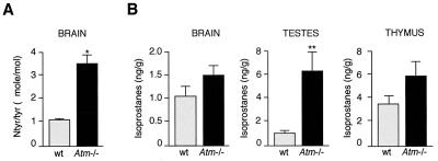
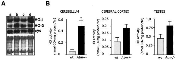
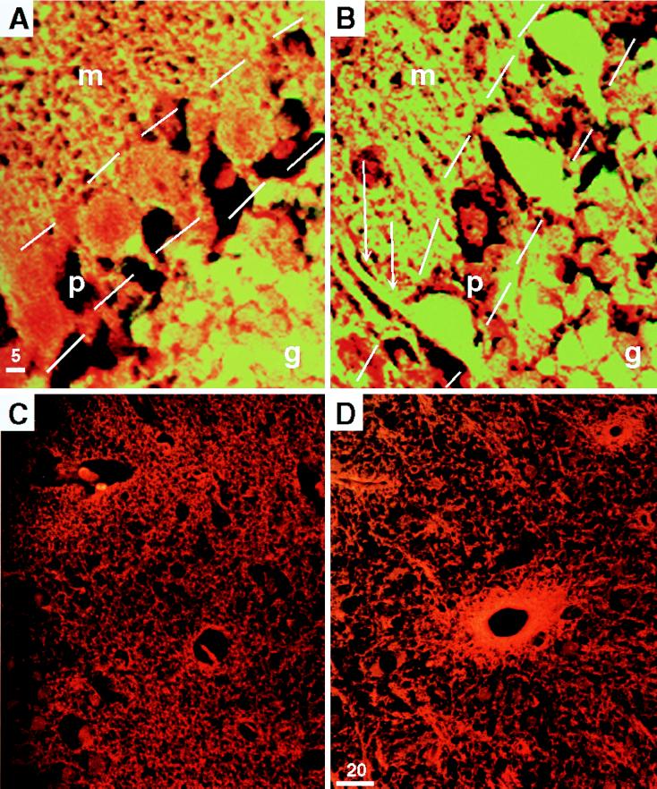
Ataxia-telangiectasia (A-T) is characterized by a markedly increased sensitivity to ionizing radiation, increased incidence of cancer, and neurodegeneration, especially of the cerebellar Purkinje cells. Ionizing radiation oxidizes macromolecules and causes tissue damage through the generation of reactive oxygen species (ROS). We therefore hypothesized that A-T is due to oxidative damage resulting from loss of function of the A-T gene product. To assess this hypothesis, we employed an animal model of A-T, the mouse with a disrupted Atm gene. We show that organs which develop pathologic changes in the Atm-deficient mice are targets of oxidative damage, and that cerebellar Purkinje cells are particularly affected. These observations provide a mechanistic basis for the A-T phenotype and lay a rational foundation for therapeutic intervention.


Figures

Figure 1
Figure 1
Proteins and lipids are covalently modified in the absence of ATM. (A) Mean ratio of nitrotyrosine to tyrosine in whole brain from 3-month-old wild-type (wt) animals (gray bar, n = 5 animals) and Atm-deficient (Atm−/−) animals (black bar, n = four animals). (B) Mean level of F2-isoprostanes from brain, testes, and thymus in wild-type (gray bars) and age-matched Atm-deficient mice (black bars). ∗, Significance with P < 0.048; ∗∗, significance with P < 0.01. Error bars represent ±SEM. Statistical analyses were performed with Fisher’s exact test.
Figure 2
Figure 2
Increased expression and activity of HO in brains from Atm-deficient mice. (A) Representative Northern blot of 10 μg of total RNA isolated from 3-month-old male littermates as follows: wild-type cerebral cortex (lane a); Atm-deficient cerebral cortex (lane b); wild-type cerebellum (lane c); and Atm-deficient cerebellum (lane d). cyc, Cyclophilin control. The bottom box is the methylene blue-stained blot for assessing RNA loading. (B) Total HO activity in the cerebellum, cerebral cortex, and testes of Atm-deficient (gray bars) and wild-type mice (black bars). Each bar represents the mean ± SEM. Statistical significance of differences between and within groups was assessed with the unpaired t test with the Scheffe correction for multiple comparisons. ∗, Significance with P < 0.05. Three animals of each genotype at 3 months of age were used for the HO activity determinations. The specific activity in wild-type cerebellum was 77 pmol of CO per mg of protein per hr; in Atm-deficient animals it was 458 pmol of CO per mg per hr (P < 0.05). The values for wild-type and Atm-deficient cortex and testes were not statistically significantly different.
Figure 3
Figure 3
Purkinje cells and cells surrounding blood vessels show increased levels of HO protein in the absence of ATM. The yellow fluorescent signal shown in A and B is from anti-HO-1 staining in a wild-type (A) and Atm-deficient (B) mouse cerebellum at high magnification (×100) to show the significant increase in staining in the Purkinje cell layer. m Indicates the molecular layer, p and outline indicate the Purkinje cell layer, and g indicates the granular cell layer. Note the intense signal in the Purkinje cell soma of the Atm-deficient brain in B. The increased staining in the molecular layer is likely caused by HO in the dendritic arbors, as the spines are well demarcated by the intense staining (double arrows). The granular cell layer (g) staining is similar in the wild-type and Atm-deficient animals. C (wild-type) and D (Atm-deficient) show the increase in bright orange signal from anti-HO-2, which is prominent in cells surrounding the vasculature of the cerebral cortex in the Atm-deficient mouse (D). (×40.)

Similar articles

Cited by

References

    1. Barlow C, Hirotsune S, Paylor R, Liyanage M, Eckhaus M, Collins F, Shiloh Y, Crawley J, Ried T, Tagle D, Wynshaw-Boris A. Cell. 1996;86:159–171. - PubMed
    1. Savitsky K, Bar-Shira A, Gilad S, Rotman G, Ziv Y, Vanagaite L, Tagle D A, Smith S, Uziel T, Sfez S, et al. Science. 1995;268:1749–1753. - PubMed
    1. Xu Y, Ashley T, Brainerd E, Bronson R, Meyn M, Baltimore D. Genes Dev. 1996;10:2411–2422. - PubMed
    1. Rotman G, Shiloh Y. Hum Mol Genet. 1998;7:1555–1563. - PubMed
    1. Brown K, Barlow C, Wynshaw-Boris A. Am J Hum Genet. 1999;64:46–50. - PMC - PubMed

Publication types