. 1999 Aug 17;96(17):9920-5.
doi: 10.1073/pnas.96.17.9920.

Stable transgene expression in rod photoreceptors after recombinant adeno-associated virus-mediated gene transfer to monkey retina

Affiliations

Stable transgene expression in rod photoreceptors after recombinant adeno-associated virus-mediated gene transfer to monkey retina

J Bennett et al. Proc Natl Acad Sci U S A. .

Abstract

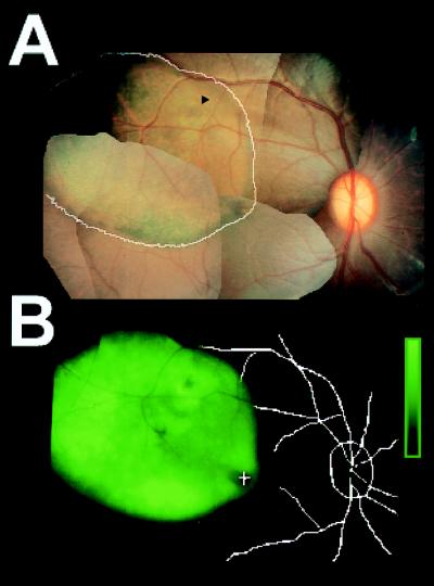
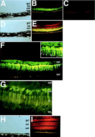
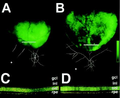
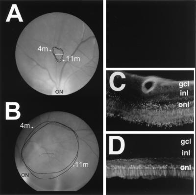
Recombinant adeno-associated virus (rAAV) is a promising vector for therapy of retinal degenerative diseases. We evaluated the efficiency, cellular specificity, and safety of retinal cell transduction in nonhuman primates after subretinal delivery of an rAAV carrying a cDNA encoding green fluorescent protein (EGFP), rAAV. CMV.EGFP. The treatment results in efficient and stable EGFP expression lasting >1 year. Transgene expression in the neural retina is limited exclusively to rod photoreceptors. There is neither electroretinographic nor histologic evidence of photoreceptor toxicity. Despite significant serum antibody responses to the vector, subretinal readministration results in additional transduction events. The findings further characterize the retinal cell tropism of rAAV. They also support the development of studies aimed ultimately at treating inherited retinal degeneration by using rAAV-mediated gene therapy.


Figures

Figure 1
Figure 1
In vivo imaging shows fluorescence localized to the site of subretinal rAAV.CMV.EGFP injection at 16 weeks. Montage of color photographs (A) and fluorescence intensity (B) in eye 1 of animal 94B-109 (Table 1). Extent of fluorescence from B is overlaid (white trace) on A. Arrowhead, injection site; Fundus landmarks (+, fovea) from A are overlaid on B. Fluorescence intensity is mapped to increasing intensities of green color; scale bar represents 2.4 log units above background.
Figure 2
Figure 2
Histologic evaluation at 6 months postinjection reveals high levels of EGFP in rod photoreceptors. Views are of retinal cross-sections from left eye of monkey 94B-102. (A, D, and H) Light microscopic views. (B and EG) Fluorescent views through an FITC filter. (C) fluorescent view through a rhodamine filter. (Inset to F) Green fluorescent protein is absent in cones (some of which are outlined or indicated with arrows). (E and I) Sections are counter-stained with propidium iodide. (H and I) Region outside of the injection site lacks EGFP; gcl, ganglion cell layer; inl, inner nuclear layer; onl, outer nuclear layer; rpe, retinal pigment epithelium; opl, outer plexiform layer; os, outer segment layer; is, inner segment layer.
Figure 3
Figure 3
Subretinal rAAV.CMV.EGFP administration to the eye contralateral to that injected with rAAV.CMV.EGFP 7 months earlier results in high levels of EGFP protein. (A and B) Fundus fluorescence 7 weeks [95C-273 eye 2 (A)] and 16 weeks [94B-051 eye 2 (B)] after readministration. Scale bar represents 2.1 log units (A) or 2.4 log units (B) of fluorescence above background. Fundus landmarks are overlaid (+, fovea). White lines in A and B indicate the regions shown histologically in C and D, respectively. gcl, ganglion cell layer; inl, inner nuclear layer; onl, outer nuclear layer; rpe, retinal pigment epithelium.
Figure 4
Figure 4
EGFP fluorescence is stable over time. Shown is the extent of in vivo fluorescence at 4 and 11 months (4 m and 11 m) postinjection in 95C-273 eye 1 (A) and 94B-051 eye 1 (B). White lines in A and B indicate the regions shown histologically in C and D, respectively. ON, optic nerve. The section shown in C was thicker than that in D (30 vs. 18 μm) to enhance detection of the weaker EGFP-labeled photoreceptors in 95C-273 eye 1. Use of the thicker section results in increased retinal autofluorescence. gcl, ganglion cell layer; inl, inner nuclear layer; onl, outer nuclear layer.

References

    1. Bundey S, Crews S J. J Med Genet. 1984;21:417–420. - PMC - PubMed
    1. Bunker C H, Berson E L, Bromley W C, Hayes R P, Roderick T H. Am J Ophthalmol. 1984;97:357–365. - PubMed
    1. Kaplan J, Bonneau D, Frezal J, Munnich A, Dufier J L. Hum Genet. 1990;85:635–642. - PubMed
    1. National Advisory Eye Council. National Institutes of Health Publication 93-3186. Bethesda, MD: National Institutes of Health; 1993.
    1. Blacharski P A. In: Fundus flavimaculatus. Newsome D A, editor. NY: Raven; 1988. pp. 135–159.

Publication types

LinkOut - more resources