. 1998 May-Jun;5(1-2):102-14.

Metamorphosis of the mushroom bodies; large-scale rearrangements of the neural substrates for associative learning and memory in Drosophila

Affiliations

Metamorphosis of the mushroom bodies; large-scale rearrangements of the neural substrates for associative learning and memory in Drosophila

J D Armstrong et al. Learn Mem. 1998 May-Jun.

Abstract

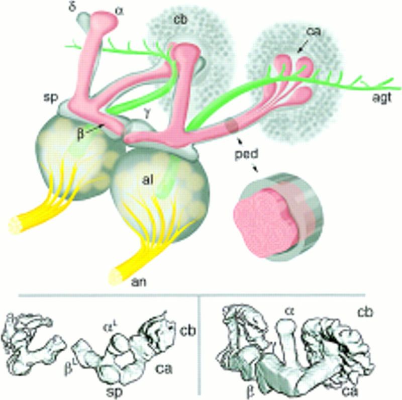
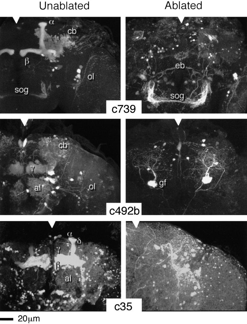
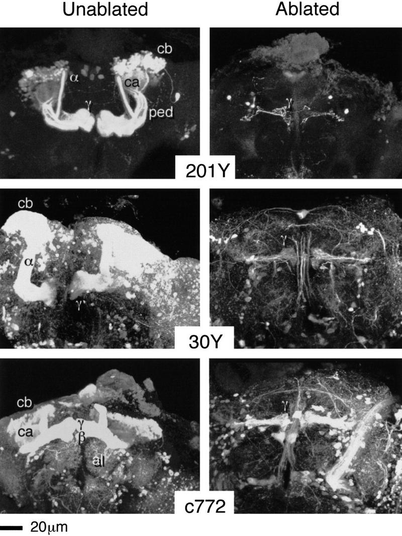
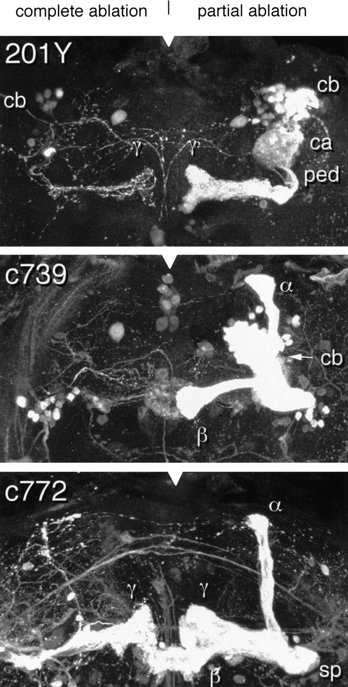
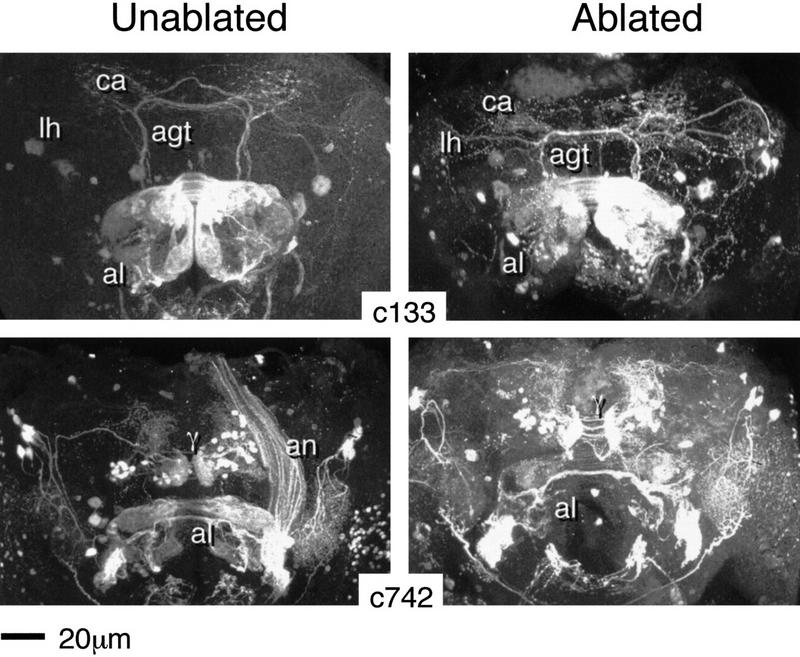
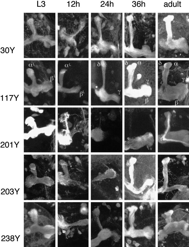
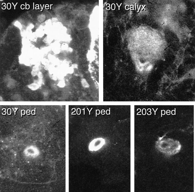
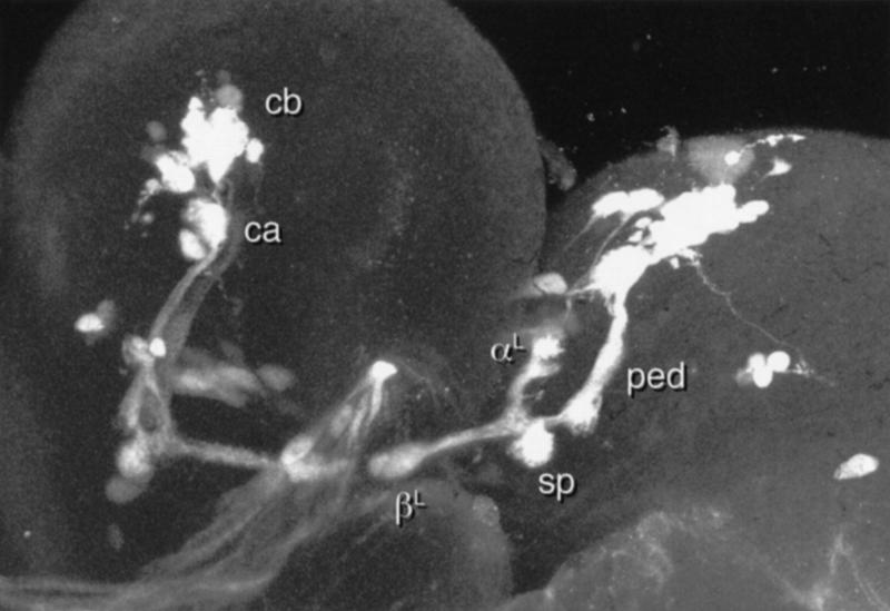
Paired brain centers known as mushroom bodies are key features of the circuitry for insect associative learning, especially when evoked by olfactory cues. Mushroom bodies have an embryonic origin, and unlike most other brain structures they exhibit developmental continuity, being prominent components of both the larval and the adult CNS. Here, we use cell-type-specific markers, provided by the P[GAL4] enhancer trap system, to follow specific subsets of mushroom body intrinsic and extrinsic neurons from the larval to the adult stage. We find marked structural differences between the larval and adult mushroom bodies, arising as the consequence of large-scale reorganization during metamorphosis. Extensive, though incomplete, degradation of the larval structure is followed by establishment of adult specific alpha and beta lobes. Kenyon cells of embryonic origin, by contrast, were found to project selectively to the adult gamma lobe. We propose that the gamma lobe stores information of relevance to both developmental stages, whereas the alpha and beta lobes have uniquely adult roles.


Figures

Figure 1
Figure 1
Drosophila mushroom bodies. (Top) Schematic representation of the adult Drosophila olfactory system. Olfactory input from sensory receptors on the antennae reaches the antennal lobes (al) via the antennal nerve (an). The glomerular arrangement of the AL is indicated. The AL also receives olfactory input from the maxillary palps (not shown). Among the outputs of the AL is the antennal glomerular tract (agt), projecting caudally toward the MB calyx (ca), and then to the lateral protocerebrum. The calyx contains the dendritic arbors of KCs and is half contained within a rind of KC cell bodies (cb). KC axons arising within the calyx project rostrally via a stalk-like structure known as the pedunculus (ped). Upon reaching the rostral margin of the central brain, the pedunculus gives rise to a spur-shaped lateral projection (sp), and to four lobes (α,β,γ,δ). β and γ project toward the midline, where they almost abut. α and δ project to the dorsal margin of the brain. MBs exhibit a clear and continuous fourfold symmetry within the cell body layer (four adjacent “clouds”), the calyx and the pedunculus (four internal tracts, each of which has a concentric organization). As they transit the pedunculus the four tracts merge, and it is their projections that form the α and β lobes. Surrounding the four peduncular tracts is a sheath of KC fibers projecting via the spur to form the γ lobe and at least some elements of the δ lobe. The inset is a schematic representation of a cross-section though the pedunculus, illustrating its internal organization. (Bottom) Surface-rendered models of late third instar larval (left) and adult (right) MBs as visualized in P{GAL4} line 201Y. The main MB structures are indicated, with the exception of the adult δ lobe, which is not encompassed by the expression pattern of line 201Y.
Figure 2
Figure 2
HU ablation of postembryonic KCs. Three-dimensional reconstructions of adult brains from the indicated P{GAL4} lines. (Notches) Approximate position of the midline in each panel. (c739) The normal expression pattern reveals the α and β lobes of the MBs, intrinsic neuronal components of the ALs (weak), the ellipsoid body—a component of the central complex (weak), together with elements of the optic lobes (ol) and the subesophageal ganglion (sog). (c492b) The normal pattern reveals large numbers of KCs within the α, β, and γ lobes (α and β less clear in this reconstruction), intrinsic neurons of the ALs (al), the giant fiber (gf, partly obscured by MB staining), and elements of the optic lobes. (c35) The normal pattern extends widely through central brain neuropil but is strongest within the MBs (α, β, γ, and δ). In each case, the ablated pattern is selectively and completely depleted for MB and AL staining. Surrounding neuropil, as far as can be determined, appears normal.
Figure 3
Figure 3
Survival of embryonic KCs. Three-dimensional reconstructions of adult brains from the indicated P{GAL4} lines. (201Y) The normal expression pattern reveals core elements of the α and β lobes, γ lobes, together with just a few other neurons. (30Y) Relatively extensive expression within the central brain includes strong MB staining (all four lobes). (c772) Again, relatively extensive expression within the central brain, strong MB staining (within all four lobes), and also a group of AL interneurons. In each case, the ablated pattern is selectively depleted for all MB staining except for a small group of KCs, different numbers in each case, projecting towards the mid-line. After ablation, so few KCs remain relative to surrounding neuropil that three-dimensional reconstructions of the entire brain tend to obscure them. The reconstructions shown are only of the lobe regions. In sections, however, surviving KCs can be followed from cell bodies of expected shape and size in the expected dorsoposterior location. They project to a very small calyx and send their axons through a narrow pedunculus.
Figure 4
Figure 4
Partial ablation–unilateral. Three-dimensional reconstructions of partially ablated MBs from the indicated lines. All lobular elements of the normal adult pattern are represented in each case. The lobes are, however, much thinner that their nonablated counterparts (cf. Figs. 2,3). The α and β lobes of 201Y are not visible in this reconstruction but can be seen in the sections from which it was created. This is consistent with the results of a similar study by Ito et al. (1997).
Figure 5
Figure 5
Ablation of MB and AL extrinsic neurons. Three-dimensional reconstructions of adult brains from the indicated P{GAL4} lines. (c133) The normal expression pattern (left) reveals two relevant neuronal types. One enters the AL via the AN, and terminates ipsi- and contralaterally within a set of dorsomedial glomeruli. The second appears to correspond to a subset of AGT fibers. These project ipsi- and contralaterally to the calyx (ca) and to the lateral protocerebrum/lateral horn (lh). The ablated pattern (right) is remarkably similar, except for a reduction in volume of the AL. (c742) The normal expression pattern (left) reveals two relevant neuronal types. One enters the AL via the AN, terminating ipsi- and contralaterally within a subset of glomeruli. The second appears to correspond to γ-exit fibers. Each hemisphere contains a group of cell bodies dorsomedial to the MB lobes that projects to a region identical in shape and position to the tip of theγ lobe. Projections to the medial protocerebrum, and via a few longer fibers to the lateral protocerebrum, are revealed. Additional and unrelated staining occurs within the lateral protocerebrum and the optic lobes. The ablated pattern (right) is remarkably similar, except for a reduction in AL volume.
Figure 6
Figure 6
Timed study of P{GAL4} expression patterns. Three-dimensional reconstructions of lobe regions from the indicated P{GAL4} lines (see also Table 2). Larval and adult patterns are in the leftmost and rightmost columns, respectively. Three representative pupal stages are shown for each line. The second column corresponds to early pupal development (∼12 hr APF), the third column to midpupal development (∼24 hr APF), and the fourth column to later pupal development (∼36 hr APF).
Figure 7
Figure 7
The late larval MBs. Selected confocal sections through the late third instar larval cell body layer (top left) and calyx (top right) of line 30Y, and through the pedunculus of 30Y and two other lines (bottom). The cell body region of 30Y appears as four clusters, each surrounding an unstained core (presumably containing the corresponding MBNbs). The junction between the calyx and the pedunculus in line 30Y appears as two tracts with unstained cores. These are continuous, with the stained and unstained components of the 30Y pedunculus. Staining of the larval pedunculus (bottom panels) appears as one or more concentric rings surrounding an unstained core.
Figure 8
Figure 8
Effect of HU ablation on the larval MBs. Three-dimensional reconstruction of the MB pattern in HU-treated late third larval instar 201Y. The ablated larval MB shows all the features of the nonablated larval pattern, albeit with much narrower lobes.

References

    1. Aceves-Pina AE, Quinn WG. Learning in normal and mutant Drosophila larvae. Science. 1979;206:93–96. - PubMed
    1. Armstrong JD, Kaiser K. The study of Drosophila brain development. In: Houdebine LM, editor. Transgenic animals—Generation and use. Harwood Academic Publishers; 1997. pp. 365–370.
    1. Brand A, Perrimon N. Targeted gene expression as a means of altering cell fates and generating dominant phenotypes. Development. 1993;118:401–415. - PubMed
    1. Brotz T M, Bochenek B, Aronstein K, ffrench-Constant R H, Borst A. γ aminobutyric acid receptor distribution in the mushroom bodies of a fly (Calliphora erythrocephala): A functional subdivision of Kenyon cells? J Comp Neurol. 1996;383:42–48. - PubMed
    1. Cambiazo V, Tully T, Krase L. Memory through metamorphosis in normal and mutant Drosophila. J Neurosci. 1994;14:68–74. - PMC - PubMed

Publication types