. 1999 Sep;19(9):5847-60.
doi: 10.1128/MCB.19.9.5847.

Functional analysis of the SIN3-histone deacetylase RPD3-RbAp48-histone H4 connection in the Xenopus oocyte

Affiliations

Functional analysis of the SIN3-histone deacetylase RPD3-RbAp48-histone H4 connection in the Xenopus oocyte

D Vermaak et al. Mol Cell Biol. 1999 Sep.

Abstract

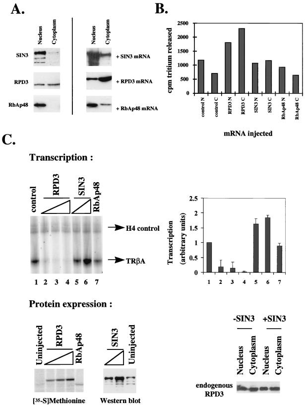
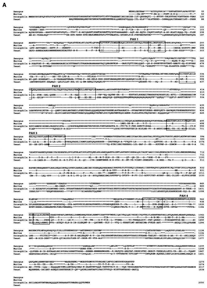
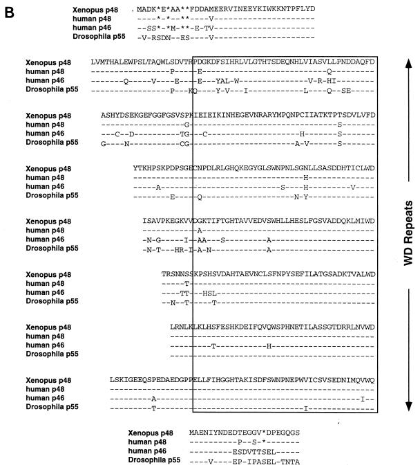
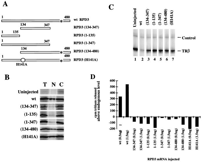
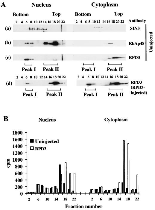
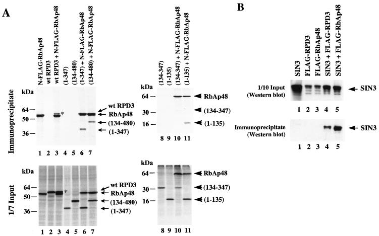
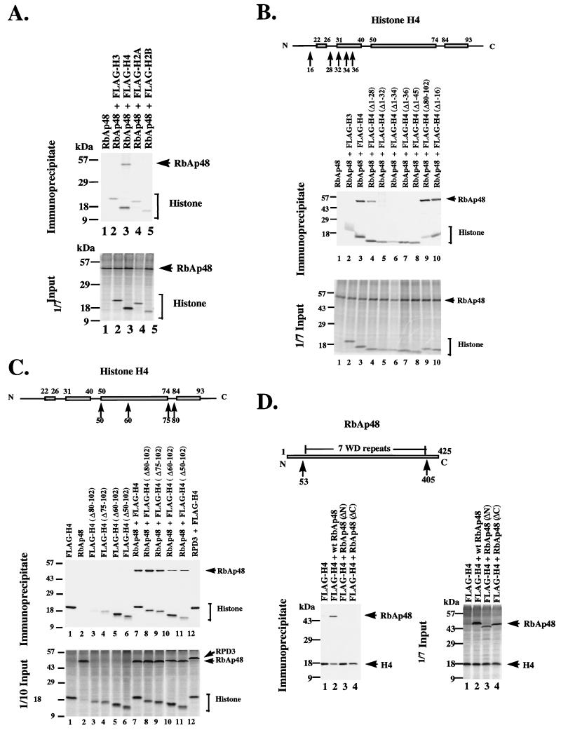
We investigated the protein associations and enzymatic requirements for the Xenopus histone deacetylase catalytic subunit RPD3 to direct transcriptional repression in Xenopus oocytes. Endogenous Xenopus RPD3 is present in nuclear and cytoplasmic pools, whereas RbAp48 and SIN3 are predominantly nuclear. We cloned Xenopus RbAp48 and SIN3 and show that expression of RPD3, but not RbAp48 or SIN3, leads to an increase in nuclear and cytoplasmic histone deacetylase activity and transcriptional repression of the TRbetaA promoter. This repression requires deacetylase activity and nuclear import of RPD3 mediated by a carboxy-terminal nuclear localization signal. Exogenous RPD3 is not incorporated into previously described oocyte deacetylase and ATPase complexes but cofractionates with a component of the endogenous RbAp48 in the oocyte nucleus. We show that RPD3 associates with RbAp48 through N- and C-terminal contacts and that RbAp48 also interacts with SIN3. Xenopus RbAp48 selectively binds to the segment of the N-terminal tail immediately proximal to the histone fold domain of histone H4 in vivo. Exogenous RPD3 may be targeted to histones through interaction with endogenous RbAp48 to direct transcriptional repression of the Xenopus TRbetaA promoter in the oocyte nucleus. However, the exogenous RPD3 deacetylase functions to repress transcription in the absence of a requirement for association with SIN3 or other targeted corepressors.


Figures

FIG. 1
FIG. 1
Nuclear distribution, transcriptional repression, and deacetylase activity of RPD3, SIN3, and RbAp48 in Xenopus oocytes. (A) Nuclear and cytoplasmic distribution of SIN3, RbAp48, and RPD3. Uninjected oocytes or oocytes which had been microinjected with SIN3, RPD3, or RbAp48 mRNA (+mRNA) were manually separated into nuclei and cytoplasm. Soluble material of five oocyte nuclei or cytoplasm equivalents was analyzed by Western blotting after SDS-PAGE. Antibodies were against SIN3, RbAp48, and RPD3. Endogenous proteins were all detected on the same Western blot. (B) Deacetylase activity of oocytes in the presence or absence of exogenous RPD3, SIN3, or RbAp48. Oocytes were uninjected (control) or microinjected with mRNA encoding RPD3, SIN3, or RbAp48, resulting in similar levels of expression of exogenous protein. Oocytes were manually dissected into nuclei (N) and cytoplasm (C), and equivalent oocyte homogenates were assayed for deacetylase activity. The error in the deacetylase assays is ±5%. (C) Exogenous RPD3 but not SIN3 or RbAp48 represses transcription from the TRβA promoter in oocytes. Oocytes were microinjected with 0.5, 1, and 1.5 ng of mRNA for RPD3 (lanes 2, 3, and 4), 1 and 1.5 ng of mRNA for SIN3 (lanes 5 and 6), or 1.5 ng of mRNA for RbAp48 (lane 7). Oocytes were maintained for 6 h to allow translation and then microinjected with 0.5 ng of double-stranded DNA for TRβA promoter. After overnight incubation, the transcript levels were analyzed by primer extension (top left). H4 serves as an internal control. The primer extension results were quantitated with a PhosphorImager and are represented graphically (top right). Protein expression was verified by labeling with [35S]methionine (RPD3 or RbAp48) or by Western blotting (SIN3) (bottom left). The distribution of endogenous RPD3 between the nucleus and cytoplasm in the presence (+SIN3) or absence (−SIN3) of exogenous SIN3 was determined by detection of RPD3 by Western blotting (bottom right).
FIG. 2
FIG. 2
Sequence comparison of xSIN3A and Xenopus RbAp48/p46 with family members from other organisms. (A) Comparison of the deduced sequence of xSIN3A with sequences of mSIN3A dSIN, and yeast SIN3A (6, 50, 74). PAH1 to PAH4 are boxed. Asterisks represent gaps and dashes identities in the sequence comparisons. (B) Manual alignment of the deduced sequence of the Drosophila and human RbAp proteins with the X. laevis sequence. WD repeats were deduced from the consensus derived from mammalian β-transducin (59, 71).
FIG. 2
FIG. 2
Sequence comparison of xSIN3A and Xenopus RbAp48/p46 with family members from other organisms. (A) Comparison of the deduced sequence of xSIN3A with sequences of mSIN3A dSIN, and yeast SIN3A (6, 50, 74). PAH1 to PAH4 are boxed. Asterisks represent gaps and dashes identities in the sequence comparisons. (B) Manual alignment of the deduced sequence of the Drosophila and human RbAp proteins with the X. laevis sequence. WD repeats were deduced from the consensus derived from mammalian β-transducin (59, 71).
FIG. 3
FIG. 3
Comparison of wt and mutant RPD3. (A) Schematic representation of RPD3 mutants. wt RPD3 is the full-length 480-aa protein (75). Numbers in parentheses indicate amino acids present in the deletion mutants. A point mutation of His to Ala at position 141 is indicated by a circle. A putative NLS (aa 438 to 444) is indicated by an asterisk. (B) Expression and nuclear localization of RPD3 constructs in oocytes. Oocytes were microinjected with 1.5 ng of mRNA encoding wt or mutant RPD3. Two oocyte equivalents of [35S]methionine-labeled protein from total oocytes (T), nuclei (N), or cytoplasm (C) were analyzed by SDS-PAGE. (C) Transcriptional repression of the TRβA promoter by wt and mutant RPD3. Oocytes were microinjected with 0.5 or 1.5 ng of mRNA, maintained for 6 h to allow translation, and then microinjected with 0.5 ng of double-stranded DNA for the TRβA promoter. After overnight incubation, the transcript levels were analyzed by primer extension. H4 serves as an internal control. (D) Histone deacetylase activity of wt and mutant RPD3 in oocyte nuclear homogenates. Nuclear oocyte extract (10 μl [one oocyte equivalent]) was assayed for histone deacetylation activity (released acetate measured as counts per minute of tritium). The level of released tritium in the absence of injected mRNA (1,100 cpm) was subtracted to yield the values shown. The error of the deacetylase assays is ±5%.
FIG. 4
FIG. 4
Distribution of nuclear and cytoplasmic RPD3, RbAp48, SIN3, and histone deacetylation activity on sucrose gradients. (A) Fractionation of nuclear and cytoplasmic endogenous RPD3, SIN3, and RbAp48 and of exogenous RPD3 on sucrose gradients. Extracts from nuclei and cytoplasm of uninjected oocytes or oocytes which had been microinjected with RPD3 mRNA were fractionated on sucrose gradients (200 oocyte equivalents per gradient). One-third of every second fraction was analyzed by Western blotting with the indicated antibodies. SIN3, RPD3, and RbAp48 were detected on the same blot for uninjected oocytes. Size markers indicated that fractions 8, 12, and 16 corresponded to 669, 443, and 150 kDa, respectively (25). (B) Expressed RPD3 cofractionates with increased histone deacetylase activity in nuclei and cytoplasm. Histone deacetylation activity of 7 μl of every second fraction was determined in uninjected and RPD3-injected nuclear and cytoplasmic fractions. Released acetate as counts per minute of tritium is indicated by numbers on the y axis. The error of the deacetylase assays is ±5%.
FIG. 5
FIG. 5
Interactions between RPD3, RbAp48, and SIN3. (A) RPD3 is coimmunoprecipitated with FLAG-tagged RbAp48. mRNAs encoding N-terminally FLAG-tagged RbAp48 and wt RPD3 or RPD3 deletion mutants were microinjected in the indicated combinations, and immunoprecipitation was carried out with anti-FLAG antibodies. RPD3 deletion mutants are indicated by amino acid numbers present in the resultant truncated proteins (Fig. 3A). One-seventh of the input extracts and all of the immunoprecipitated [35S]methionine-labeled proteins were analyzed by SDS-PAGE. Full-length RPD3 protein is marked on the upper right-hand corner with an asterisk. (B) SIN3 coimmunoprecipitates with RPD3 and with RbAp48. mRNAs encoding SIN3 and FLAG-tagged RPD3 or RbAp48 were injected as indicated above each lane. One-tenth of the input extracts and anti-FLAG-immunoprecipitated proteins were analyzed by Western blotting with anti-SIN3 antibodies.
FIG. 6
FIG. 6
Interaction of RbAp48 with core histones. (A) RbAp48 interacts specifically with core histone H4. mRNAs encoding FLAG-tagged core histones were coinjected with RbAp48 mRNA as indicated, and immunoprecipitation was carried out with anti-FLAG antibodies. One-seventh of the input extracts and immunoprecipitated [35S]methionine-labeled proteins were analyzed by SDS-PAGE. (B) RbAp48 interacts with a region in the N-terminal helix of the histone fold motif of H4. Details are as for panel A except that mRNAs encoding FLAG-tagged full-length and N-terminally deleted H4 were coinjected with RbAp48 mRNA as indicated. Deleted amino acids are preceded by Δ. (C) The C-terminal region of H4 up to aa 50 is dispensable for the interaction with RbAp48. Details are as for panel A except that mRNAs encoding FLAG-tagged full-length and C-terminally deleted H4 were coinjected with RbAp48 mRNA as indicated. (D) Removal of either the N- or C-terminal region of RbAp48 abolishes interaction with H4. Details are as for panel A except that mRNA encoding FLAG-tagged full-length H4 was coinjected with full-length and N- or C-terminally deleted RbAp48 mRNA [RbAp48(ΔN) or RbAp48(ΔC)] as indicated. For comparison, the first WD repeat is from aa 55 to 94 and the seventh and last WD repeat is from aa 363 to 403.

References

    1. Alland L, Muhle R, Hou H, Jr, Potes J, Chin L, Schreiber-Agus N, De Pinho R A. Role of NCoR and histone deacetylase in Sin3-mediated transcriptional and oncogenic repression. Nature. 1997;387:49–55. - PubMed
    1. Almouzni G, Wolffe A P. Replication-coupled chromatin assembly is required for the repression of basal transcription in vivo. Genes Dev. 1993;7:2033–2047. - PubMed
    1. Almouzni G, Wolffe A P. Nuclear assembly, structure, and function: the use of Xenopus in vitro systems. Exp Cell Res. 1993;205:1–15. - PubMed
    1. Almouzni G, Mechali M. Assembly of spaced chromatin by DNA synthesis in extracts from Xenopus eggs. EMBO J. 1988;7:664–672. - PMC - PubMed
    1. Annunziato A T. Histone acetylation during chromatin replication and nucleosome assembly. Nucleus. 1995;1:31–58.

Publication types

MeSH terms

Associated data

LinkOut - more resources