. 1999 Sep;19(9):5923-9.
doi: 10.1128/MCB.19.9.5923.

NF-kappaB induces expression of the Bcl-2 homologue A1/Bfl-1 to preferentially suppress chemotherapy-induced apoptosis

Affiliations

NF-kappaB induces expression of the Bcl-2 homologue A1/Bfl-1 to preferentially suppress chemotherapy-induced apoptosis

C Y Wang et al. Mol Cell Biol. 1999 Sep.

Abstract

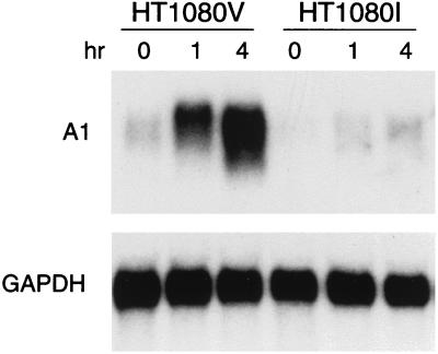
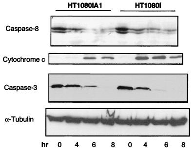
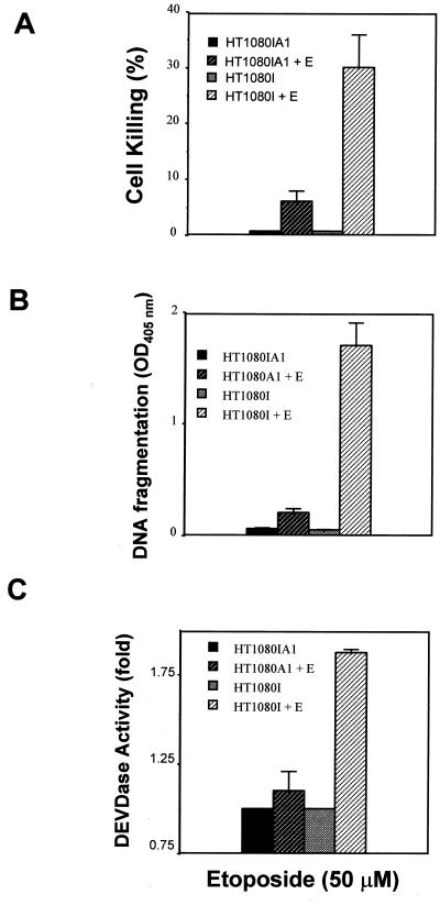
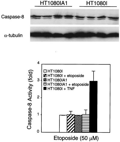
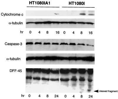
Recent evidence indicates that the transcription factor NF-kappaB is a major effector of inducible antiapoptotic mechanisms. For example, it was shown that NF-kappaB activation suppresses the activation of caspase 8, the apical caspase in tumor necrosis factor (TNF) receptor family signaling cascades, through the transcriptional regulation of certain TRAF and IAP proteins. However, it was unknown whether NF-kappaB controls other key regulatory mechanisms in apoptosis. Here we show that NF-kappaB activation suppresses mitochondrial release of cytochrome c through the activation of the Bcl-2 family member A1/Bfl-1. The restoration of A1 in NF-kappaB null cells diminished TNF-induced apoptosis by reducing the release of proapoptotic cytochrome c from mitochondria. In addition, A1 potently inhibited etoposide-induced apoptosis by inhibiting the release of cytochrome c and by blocking caspase 3 activation. Our findings demonstrate that A1 is an important antiapoptotic gene controlled by NF-kappaB and establish that the prosurvival function of NF-kappaB can be manifested at multiple levels.


Figures

FIG. 1
FIG. 1
A1/Bfl-1 expression is induced by TNF through the activation of NF-κB. HT1080V and HT1080I cells were treated with TNF-α (20 ng/ml) for the indicated time. Total RNA was extracted with Trizol reagent. Northern blots were performed as described in Materials and Methods. The filter was probed with 32P-labeled human A1 cDNA probe. For the internal control, the blot was stripped and reprobed with 32P-labeled GAPDH (glyceraldehyde-3-phosphate dehydrogenase) cDNA probe.
FIG. 2
FIG. 2
A1 expression in HT1080I cells partially inhibits TNF-induced apoptosis. (A) Detection of stable HT1080I transfectants expressing A1/Bfl-1. HT1080I cells were transfected with pcDNA3-Flag-A1 vector or control empty vector and selected with G418 (600 μg/ml) for 2 weeks. The clones were analyzed by monoclonal antibody against Flag epitope. The five stable clones which expressed similar levels of A1 protein were pooled (HT1080IA1). Lane 1 and 2 represent the stable HT1080I cells expressing A1; lane 3 represents the stable control clones expressing G418-resistant marker (HT1080I). (B and C) A1 partially inhibits TNF-induced apoptosis. The stable cell clones used for panel A were treated with TNF (20 ng/ml) for the indicated times. Cell viability was determined by trypan blue exclusion. The supernatants from the 14-h time point of TNF treatment were collected and measured by cell death ELISA. The results represent the mean values from three independent experiments. (D) A1 inhibits TNF-induced DEVDase activity. The stable cell clones used for panel A were treated with TNF (20 ng/ml) for 6 h. Cells were lysed in hypotonic buffer (see Materials and Methods), and 300-μg aliquots of extracts were incubated with DEVD-pNA (100 μM) substrate for 2 h at 37°C. The reaction was measured with a plate reader by determining the OD405. The results represent the mean values from three independent experiments.
FIG. 3
FIG. 3
A1 partially inhibits TNF-induced cytochrome c release from mitochondria and inhibits caspase 3 processing. HT1080IA1 and HT1080I cells were treated with TNF (20 ng/ml) for the indicated times. To detect the release of cytochrome c, cytosolic proteins were extracted and separated by SDS–15% polyacrylamide gel electrophoresis. The blot was probed with monoclonal antibody to cytochrome c (1:1,000). For detecting caspase 8 and caspase 3 processing, whole-cell extracts were prepared and immunoblotted with monoclonal antibodies to caspase 8 (1:1,000) and caspase 3 (1:500). For the internal control, the blots were stripped and reprobed with antibody to α-tubulin (1:2,000).
FIG. 4
FIG. 4
A1/Bfl-1 expression is induced by etoposide through the activation of NF-κB. (A) Etoposide induces the nuclear translocation of NF-κB in HT1080V cells but not HT1080I cells. Cells were treated with etoposide (50 μM) for the indicated time. EMSAs were performed as described in Materials and Methods. (B) A1 gene expression is induced by etoposide. Cells were treated with etoposide (50 μM) for the indicated time. Northern blot analyses were performed as described for Fig. 1.
FIG. 5
FIG. 5
A1 inhibits etoposide-induced apoptosis. (A and B) HT1080IA1 and HT1080I cells were treated with etoposide (E; 50 μM) for 24 h. The cell viability and cell death ELISAs were performed as described for Fig. 2B and C, respectively. (C) A1 inhibits etoposide-induced DEVDase activity. Cells were treated with etoposide (50 μM) for 16 h, and DEVDase assays were performed as described for Fig. 2D. The results represent the average values from three independent experiments.
FIG. 6
FIG. 6
Caspase 8 activity is not induced by etoposide. HT1080IA1 and HT1080I cells were treated with etoposide (50 μM) for 24 h. The whole-cell extracts were probed with a monoclonal antibody against caspase 8 as described for Fig. 3. The lanes are as shown in Fig. 7. For detecting caspase 8 activity, cells were treated with etoposide (50 μm) for 24 h and then lysed in hypotonic buffer as described for Fig. 2, and 300-μg aliquots of protein extracts were incubated with IETD-pNA substrate (100 μM) for 2 h at 37°C. The reaction was measured with a plate reader by determining the OD405. The results represent the average values from three independent experiments.
FIG. 7
FIG. 7
A1 potently inhibits etoposide-induced cytochrome c release. HT1080IA1 and HT1080I cells were treated with etoposide (50 μM) for the indicated times. Cytosolic proteins were extracted and probed with a monoclonal antibody against cytochrome c. For detecting caspase 3 and DFF45 processing, the whole-cell extracts were prepared as described for Fig. 3. For internal control, the blots were stripped and reprobed with monoclonal antibody to α-tubulin.

References

    1. Adams J, Cory S. The Bcl-2 protein family: arbiters of cell survival. Science. 1998;281:1322–1326. - PubMed
    1. Ashkenazi A, Dixit V. Death receptors: signaling and modulation. Science. 1998;281:1305–1308. - PubMed
    1. Baeuerle P A, Baltimore D. NF-κB: ten years after. Cell. 1996;87:13–20. - PubMed
    1. Baker S, Reddy E P. Modulation of life and death by the TNF receptor superfamily. Oncogene. 1998;25:3261–3270. - PubMed
    1. Baldwin A S. The NF-κB and IκB proteins: new discoveries and insights. Annu Rev Immunol. 1996;14:649–681. - PubMed

Publication types

MeSH terms

LinkOut - more resources