. 1999 Sep;19(9):6041-7.
doi: 10.1128/MCB.19.9.6041.

Cell cycle progression and proliferation despite 4BP-1 dephosphorylation

Affiliations

Cell cycle progression and proliferation despite 4BP-1 dephosphorylation

S O Marx et al. Mol Cell Biol. 1999 Sep.

Abstract

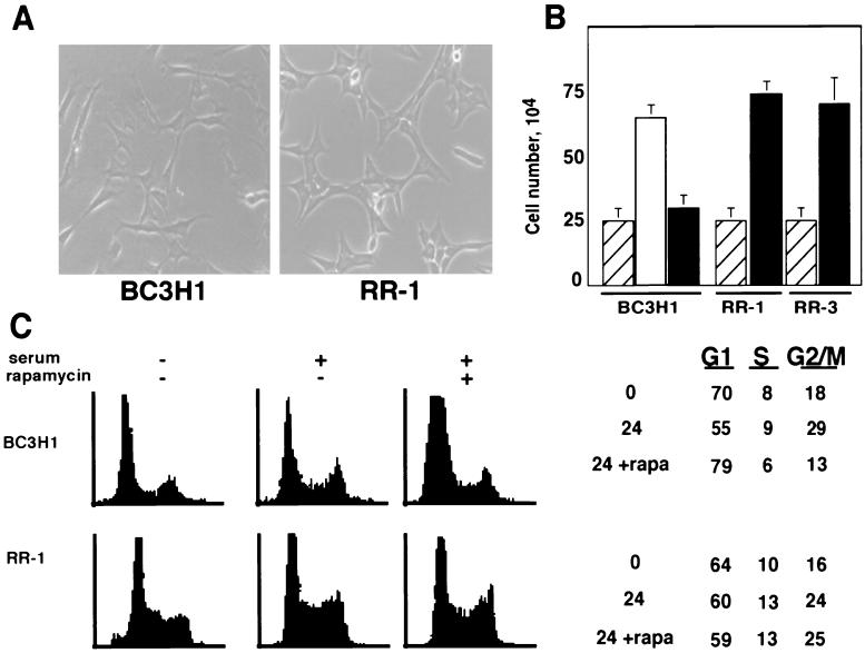
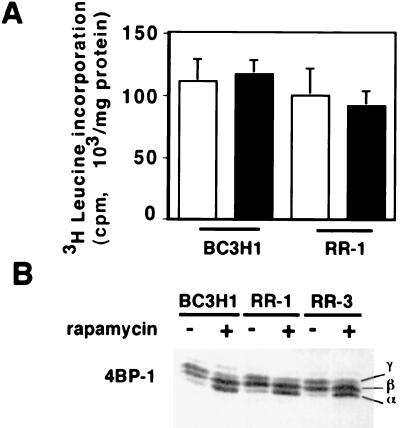
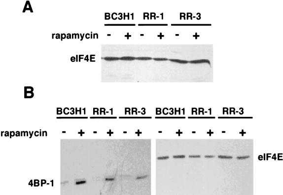
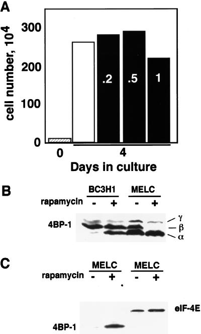
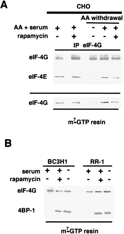
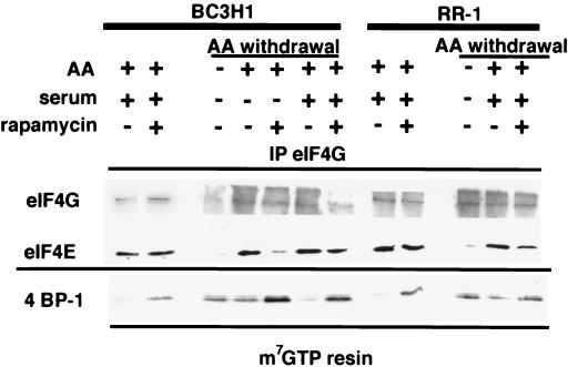
Proliferation and cell cycle progression in response to growth factors require de novo protein synthesis. It has been proposed that binding of the eukaryotic translation initiation factor 4E (eIF-4E) to the inhibitory protein 4BP-1 blocks translation by preventing access of eIF-4G to the 5' cap of the mRNA. The signal for translation initiation is thought to involve phosphorylation of 4BP-1, which causes it to dissociate from eIF-4E and allows eIF-4G to localize to the 5' cap. It has been suggested that the ability of the macrolide antibiotic rapamycin to inhibit 4BP-1 phosphorylation is responsible for the potent antiproliferative property of this drug. We now show that rapamycin-resistant cells exhibited normal proliferation despite dephosphorylation of 4BP-1 that allows it to bind to eIF-4E. Moreover, despite rapamycin-induced dephosphorylation of 4BP-1, eIF-4E-eIF-4G complexes (eIF-4F) were still detected. In contrast, amino acid withdrawal, which caused a similar degree of 4BP-1 dephosphorylation, resulted in dissociation of the eIF-4E-eIF-4G complex. Thus, 4BP-1 dephosphorylation is not equivalent to eIF-4E inactivation and does not explain the antiproliferative property of rapamycin.


Figures

FIG. 1
FIG. 1
Growth characteristics of RR cells. (A) The morphologies and sizes of proliferating parental (BC3H1) and RR-1 cells are similar. (B) Rapamycin inhibited cell growth in the parental cells but not in two RR cell lines, RR-1 and RR-3. Hatched bars show the numbers of cells that were plated; cell number was determined after 48 h. Growth was significantly inhibited for rapamycin-treated cells (100 nM, black bars) compared to untreated BC3H1 cells (white bar). Data are averages of triplicate samples + standard deviations. (C) Flow cytometry of parental and RR-1 cells. Cells were serum starved in DMEM plus 1% FBS for 24 h, followed by stimulation with 20% FBS. Cells were harvested after 24 h and stained with propidium iodide. Analysis was based upon results from a minimum of 15,000 cells. Parental cells treated with rapamycin (100 nM) arrested in G1/S; RR-1 cells showed no rapamycin-induced inhibition of cell cycle progression.
FIG. 2
FIG. 2
Generalized protein synthesis is unaffected by rapamycin, but 4BP-1 is inhibited in parental and RR cells. (A) Parental BC3H1 and RR-1 cells (104) were cultured in DMEM plus 20% FBS for 48 h in 12-well dishes; 1 μM rapamycin (black bars) or vehicle (white bars) was added to the appropriate wells when the cells were pulsed with [3H]leucine (1 μCi). Incorporation of [3H]leucine was measured by precipitation with trichloroacetic acid. Rapamycin had no significant effect on protein synthesis. Data are representative of three experiments. (B) Parental BC3H1, RR-1, and RR-3 cells were cultured in 20% FBS; 100 nM rapamycin or vehicle was added to the cultures, and lysates were prepared after 45 min. Cellular lysates (100 μg) were analyzed by immunoblotting with an anti-4BP-1 (anti-PHAS-I) antiserum. α, β, and γ are arbitrary designations of bands representing the phosphorylated forms of 4BP-1 (23).
FIG. 3
FIG. 3
Rapamycin increases complex formation between 4BP-1 and eIF-4E in parental and RR cells. (A) Cell extracts (100 μg) were analyzed by immunoblotting with anti-eIF-4E antibody. eIF-4E protein levels were equivalent in parental BC3H1 and RR cells. (B) Binding of 4BP-1 to eIF-4E (already bound to m7-GTP–Sepharose resin) was determined. m7-GTP–Sepharose was incubated with 130 μg of cellular extract for 1 h at 4°C. The resin was washed, size fractionated by SDS-PAGE, transferred to nitrocellulose, and blotted with anti-4BP-1 or anti-eIF-4E antibody.
FIG. 4
FIG. 4
MELC are rapamycin resistant and demonstrate rapamycin-induced 4BP-1 dephosphorylation and increased 4BP-1–eIF-4E complex formation. (A) MELC (10 × 104 cells; hatched bar) were treated with either a vehicle (white bar) or increasing concentrations of rapamycin (black bars [numbers are micromolar concentrations]). Cells were counted at 4 days. These data are representative of two experiments. (B) Cell extracts were analyzed by immunoblotting with an anti-4BP-1 antibody. Rapamycin (0.2 μM) treatment for 1 h induced dephosphorylation of 4BP-1. α, β, and γ are arbitrary designations of bands representing the phosphorylated forms of 4BP-1 (23). Data are representative of three experiments. (C) Binding of 4BP-1 to eIF-4E (bound to m7-GTP resin) was determined in the presence or absence of rapamycin (0.2 μM). Cellular extracts were incubated with m7-GTP resin, size fractionated by SDS-PAGE, transferred to nitrocellulose, and immunoblotted with anti-4BP-1 and anti-eIF-4E antibodies. Rapamycin increased the binding of 4BP-1 to eIF-4E.
FIG. 5
FIG. 5
Rapamycin does not prevent serum-induced association of eIF-4E–eIF-4G in CHO, BC3H1, and RR cells. (A) CHO cells were grown in DMEM plus 10% FBS and treated with either a vehicle or rapamycin for 24 h. In parallel experiments, CHO cells were placed in Ham’s F-12 medium without serum for 16 h. Cells were washed and amino acid deprived in Earle’s salt solution for 1 h. Cells were stimulated with Ham’s F-12 medium plus 10% FBS following pretreatment with either a vehicle or rapamycin (1 μM). Cell lysates were prepared as described in Materials and Methods. Cellular extracts (100 μg) were incubated with m7-GTP resin at 4°C in duplicate, washed, size fractionated on an SDS–8% polyacrylamide gel, transferred to nitrocellulose, and immunoblotted with anti-eIF-4G antibody (the nitrocellulose was also incubated with anti-eIF-4E antibody to demonstrate equal uptake on the resin [data not shown]). Cellular extracts (100 μg) were immunoprecipitated with anti-eIF-4G antibody, bound with protein A-Sepharose beads, size fractionated on an SDS–11.5% polyacrylamide gel, transferred to nitrocellulose, and immunoblotted with either anti-eIF-4E or anti-eIF-4G antibodies. Amino acid (AA) withdrawal significantly inhibited eIF-4E–eIF-4G complex formation. Rapamycin had no effect on serum-stimulated eIF-4E–eIF-4G association. Data are representative of three similar experiments. (B) BC3H1 and RR-1 cells were grown in DMEM plus 20% FBS. Experiments were performed as described above (in duplicate) to determine the binding of eIF-4G and 4BP-1 to eIF-4E bound to m7-GTP–Sepharose resin. Size fractionation was performed by SDS-PAGE with either 8% (eIF-4G) or 13% (4BP-1) polyacrylamide. Rapamycin (1 μM) was added to the medium 24 h prior to cell lysis. Rapamycin and serum withdrawal (cells maintained in DMEM alone) for 1 h caused increased association of 4BP-1 with the m7-GTP resin in BC3H1 and RR cells. However, complex formation between eIF-4E and eIF-4G persisted despite rapamycin (1 μM) treatment for 24 h.
FIG. 6
FIG. 6
Differential regulation of the eIF-4E–eIF-4G complex by rapamycin and amino acid withdrawal in BC3H1 and RR-1 cells. BC3H1 and RR-1 cells were cultured in DMEM plus 20% FBS and treated with rapamycin (1 μM). In amino acid (AA) withdrawal experiments, cells were placed in Earle’s balanced salt solution for 1 h. Cells were pretreated with rapamycin (1 μM) for 15 minutes and then stimulated for 30 min either with amino acids or with 20% FBS plus amino acids. Cellular extracts were prepared. eIF-4G and eIF-4E were coimmunoprecipitated with an anti-eIF-4G antibody and immunoblotted with either anti-eIF-4G or anti-eIF-4E antibody. In addition, the same extracts were used in experiments performed as described above (in duplicate) to determine the binding of 4BP-1 to eIF-4E (already bound to m7-GTP–Sepharose resin).

References

    1. Beretta L, Gingras A-C, Svitkin Y V, Hall M N, Sonenberg N. Rapamycin blocks the phosphorylation of 4E-BP1 and inhibits cap-dependent initiation of translation. EMBO J. 1996;15:658–664. - PMC - PubMed
    1. Blackshear P J, Stumpo D J, Carballo E, Lawrence J C. Disruption of the gene encoding the mitogen-regulated translational modulator PHAS-I in mice. J Biol Chem. 1997;272:31510–31514. - PubMed
    1. Blommaart E F, Luiken J J, Blommaart P J, van Woerkom G M, Meijer A J. Phosphorylation of ribosomal protein S6 is inhibitory for autophagy in isolated rat hepatocytes. J Biol Chem. 1995;270:2320–2326. - PubMed
    1. Brown E, Albers T, Shin T, Ichikawa K, Keith C, Lane W, Schreiber S. A mammalian protein targeted by G1-arresting rapamycin complex. Nature (London) 1994;369:756–758. - PubMed
    1. Brown E J, Beal P A, Keith C T, Chen J, Shin T B, Schreiber S L. Control of p70 S6 kinase by kinase activity of FRAP in vivo. Nature (London) 1995;377:441–446. - PubMed

Publication types

MeSH terms

LinkOut - more resources