. 1999 Sep;19(9):6057-64.
doi: 10.1128/MCB.19.9.6057.

The strength of interaction at the Raf cysteine-rich domain is a critical determinant of response of Raf to Ras family small GTPases

Affiliations

The strength of interaction at the Raf cysteine-rich domain is a critical determinant of response of Raf to Ras family small GTPases

T Okada et al. Mol Cell Biol. 1999 Sep.

Abstract

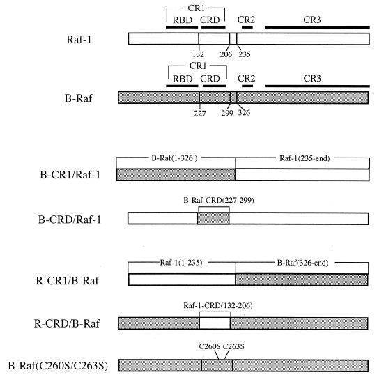
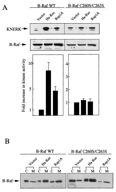
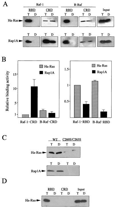
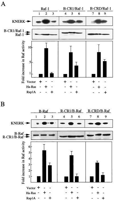
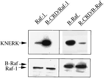
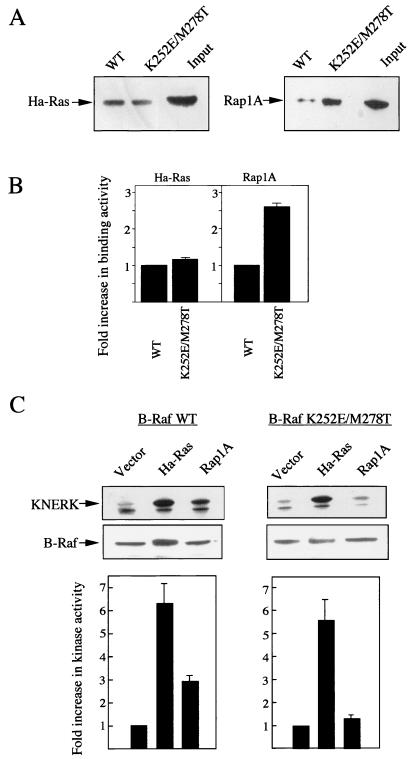
To be fully activated at the plasma membrane, Raf-1 must establish two distinct modes of interactions with Ras, one through its Ras-binding domain and the other through its cysteine-rich domain (CRD). The Ras homologue Rap1A is incapable of activating Raf-1 and even antagonizes Ras-dependent activation of Raf-1. We proposed previously that this property of Rap1A may be attributable to its greatly enhanced interaction with Raf-1 CRD compared to Ras. On the other hand, B-Raf, another Raf family member, is activatable by both Ras and Rap1A. When interactions with Ras and Rap1A were measured, B-Raf CRD did not exhibit the enhanced interaction with Rap1A, suggesting that the strength of interaction at CRDs may account for the differential action of Rap1A on Raf-1 and B-Raf. The importance of the interaction at the CRD is further supported by a domain-shuffling experiment between Raf-1 and B-Raf, which clearly indicated that the nature of CRD determines the specificity of response to Rap1A: Raf-1, whose CRD is replaced by B-Raf CRD, became activatable by Rap1A, whereas B-Raf, whose CRD is replaced by Raf-1 CRD, lost its response to Rap1A. Finally, a B-Raf CRD mutant whose interaction with Rap1A is selectively enhanced was isolated and found to possess the double mutation K252E/M278T. B-Raf carrying this mutation was not activated by Rap1A but retained its response to Ras. These results indicate that the strength of interaction with Ras and Rap1A at its CRD may be a critical determinant of regulation of the Raf kinase activity by the Ras family small GTPases.


Figures

FIG. 1
FIG. 1
Schematic representation of the structures of Raf-1, B-Raf, and their chimeric constructs. The top two horizontal bars represent the structures of Raf-1 and B-Raf protein, on which three conserved regions (CR1, CR2, and CR3) and the two subregions (RBD and CRD) in CR1 are indicated. See the text for the definition of these regions and subregions. The structures of chimeric constructs of Raf-1 and B-Raf used in this study are also shown. Numbers below the bars represent the amino acid positions of restriction cleavage sites which were used for exchanging various domains between Raf-1 and B-Raf.
FIG. 2
FIG. 2
Ras- and Rap1A-dependent activation and membrane translocation of B-Raf and B-Raf(C260S/C263S). (A) pH8-FLAG-B-Raf (wild type [WT]) or pH8-FLAG-B-Raf(C260S/C263S) (0.5 μg of each) was cotransfected with either pEF-BOS-Ha-RasVal-12, pSRα-Rap1AVal-12, or pEF-BOS (3 μg of each) into COS7 cells. FLAG–B-Raf proteins were immunoprecipitated from the total cellular extract and examined for induction of phosphorylation of GST-KNERK in the presence of GST-MEK as described in Materials and Methods. The upper panel shows autoradiograms of phosphorylated GST-KNERK. The intensity of the KNERK bands was quantified with a BAS2000 bioimaging analyzer (lower panel) and expressed as fold increase with respect to the cells cotransfected with pEF-BOS. Immunoblot detection of B-Raf in the extracts is shown in the middle panel. The data shown are the means of three independent experiments. Standard deviations are indicated as error bars. (B) The transfected cells were homogenized and separated into cytosol (C) and membrane (M) fractions. B-Raf proteins present in the two fractions were detected by Western immunoblotting with anti-B-Raf antibody.
FIG. 3
FIG. 3
Association of Ha-Ras and Rap1A with various subfragments of Raf-1, B-Raf, and B-Raf(C260S/C263S). (A) Ha-Ras and Rap1A (10 pmol of each) were loaded with GTPγS (T) or GDPβS (D) and examined for association with 25 pmol of MBP–Raf-1(51-131) (Raf-1 RBD) or MBP–B-Raf(144-226) (B-Raf RBD) and with 100 pmol of MBP–Raf-1(132-206) (Raf-1 CRD) or MBP–B-Raf(227-299) (B-Raf CRD), which were immobilized on amylose resin, as described in Materials and Methods. The bound Ha-Ras and Rap1A were fractioned by SDS-PAGE (12% gel) and subjected to Western immunoblotting with anti-Ha-Ras and anti-Rap1A antibodies, respectively. One-tenth of the amount of Ha-Ras or Rap1A used for the binding reactions was applied on the same gel (Input). All Western blots were exposed to X-ray film for the same period. The sensitivities of anti-Ha-Ras and anti-Rap1A antibodies were almost equal, as observed before (15, 16). (B) Ha-Ras and Rap1A (20 pmol for CRD binding and 10 pmol for RBD binding) were loaded with [γ-35S]GTPγS and examined for association with 60 pmol of MBP fusion proteins immobilized on amylose resin as described in Materials and Methods. The results shown are the means of three independent experiments performed in duplicate and expressed as fold differences where the amount of Ha-Ras bound to Raf-1 CRD (1.8 pmol) (left) or to Raf-1 RBD (3.8 pmol) (right) is defined as 1. Standard deviations are indicated as error bars. (C) In vitro binding reactions carried out as for panel A by using 100 pmol each of MBP–B-Raf(227-299) (wild type [WT]) and MBP–B-Raf(C260S/C263S). (D) Ten picomoles of posttranslationally unmodified Ha-Ras was loaded with GTPγS (T) or GDPβS (D) and incubated with 25 pmol of MBP–B-Raf(144-226) (B-Raf RBD) or 100 pmol of MBP–B-Raf(227-299) (B-Raf CRD) as described for panel A.
FIG. 4
FIG. 4
Ras- and Rap1A-dependent kinase activities of Raf-1, B-Raf, and their chimeric constructs. (A) pH8-FLAG-Raf-1, pH8-FLAG-B-CR1/Raf-1, or pH8-FLAG-B-CRD/Raf-1 (3 μg of each) was cotransfected into COS7 cells with either pEF-BOS-Ha-RasVal-12, pSRα-Rap1AVal-12, or pEF-BOS (3 μg of each). The kinase activities of Raf immunoprecipitated from the total cellular extracts were measured as described in Materials and Methods. The upper panel shows autoradiograms of the phosphorylated GST-KNERK. The intensity of the KNERK bands was quantified as for Fig. 2A (lower panel). The amounts of FLAG–Raf-1 and its chimeras in the extracts were measured by immunoblotting with anti-Raf-1 antibody (middle panel). (B) pH8-FLAG-B-Raf, pH8-FLAG-R-CR1/B-Raf, or pH8-FLAG-R-CRD/B-Raf (0.5 μg of each) was cotransfected into COS7 cells with either pEF-BOS-Ha-RasVal-12, pSRα-Rap1AVal-12, or pEF-BOS (3 μg of each), and their kinase activities were measured as described for panel A. The upper panel shows the autoradiograms of the phosphorylated GST-KNERK. The intensity of the KNERK bands was quantified as for Fig. 2A (lower panel). The amounts of FLAG–B-Raf and its chimeras in the extracts were measured by immunoblotting with anti-B-Raf antibody (middle panel). The data shown are the means of two independent experiments performed in duplicate. Standard deviations are indicated as error bars.
FIG. 5
FIG. 5
Rap1A-dependent activation of Raf-1, B-Raf, and their chimeras translocated to the membrane. pH8-FLAG-Raf-1, pH8-FLAG-B-CRD/Raf-1, pH8-FLAG-B-Raf, or pH8-FLAG-R-CRD/B-Raf (3 μg of each) was cotransfected into COS7 cells with 3 μg of pSRα-Rap1AVal-12. The transfected cells were homogenized and separated into cytosol and membrane fractions. The kinase activities of Raf immunoprecipitated from the membrane extracts were measured as described in Materials and Methods. The upper panel shows the autoradiograms of the phosphorylated GST-KNERK. The amounts of FLAG–Raf-1 or FLAG–B-Raf and their chimeras in the membrane extracts were measured by immunoblotting with anti-Raf-1 antibody or anti-B-Raf antibody (lower panel). Three independent experiments were performed with similar results.
FIG. 6
FIG. 6
Effects of a K252E/M278T mutation on the interaction of B-Raf-CRD with, and activation of B-Raf by, Ras and Rap1A. (A) MBP–B-Raf(227-299) (wild type [WT]) and MBP–B-Raf (K252E/M278T) were examined for in vitro association with Ha-Ras or Rap1A as described in the legend to Fig. 3. The bound Ha-Ras and Rap1A were fractioned by SDS-PAGE (12% gel) and subjected to Western immunoblotting with anti-Ha-Ras and anti-Rap1A antibodies, respectively. One-tenth of the amount of Ha-Ras or Rap1A used for the binding reactions were applied on the same gel (Input). (B) Ha-Ras and Rap1A (20 pmol of each) were loaded with [γ-35S]GTPγS and examined for association with 60 pmol each of MBP–B-Raf(227-299) (WT) and MBP–B-Raf(K252E/M278T) as described in Materials and Methods. The results shown are the means of three independent experiments performed in duplicate and expressed as fold increases over the binding activities of wild-type B-Raf CRD (1.5 pmol for Ha-Ras and 1.2 pmol for Rap1A). Standard deviations are indicated as error bars. (C) pH8-FLAG-B-Raf or pH8-FLAG-B-Raf(K252E/M278T) (0.5 μg of each) was cotransfected into COS7 cells with either pEF-BOS-Ha-RasVal-12, pSRα-Rap1AVal-12, or pEF-BOS (3 μg of each). The kinase activities of B-Raf immunoprecipitated from the total cellular extracts were measured as described in Materials and Methods. The upper panel shows the autoradiograms of the phosphorylated GST-KNERK. The intensity of the KNERK bands was quantified as for Fig. 2A (lower panel). The amounts of FLAG–B-Raf in the extracts were measured by immunoblotting with anti-B-Raf antibody (middle panel). The data shown are the means of three independent experiments. Standard deviations are indicated as error bars.

References

    1. Ayruch J. Insulin signal transduction through protein kinase cascades. Mol Cell Biochem. 1998;182:31–48. - PubMed
    1. Bosch E, Cherwinski H, Peterson D, McMahon M. Mutations of critical amino acids affect the biological and biochemical properties of oncogenic A-Raf and Raf-1. Oncogene. 1997;15:1021–1033. - PubMed
    1. Brtva T R, Drugan J K, Ghosh S, Terrell R S, Campbell-Burk S, Bell R M, Der C J. Two distinct Raf domains mediate interaction with Ras. J Biol Chem. 1995;270:9809–9812. - PubMed
    1. Campbell S L, Khosravi-Far R, Rossman K L, Clark G J, Der C J. Increasing complexity of Ras signaling. Oncogene. 1998;17:1395–1413. - PubMed
    1. Chuang E, Barnard D, Hettich L, Zhang X-F, Avruch J, Marshall M S. Critical binding and regulatory interactions between Ras and Raf occur through a small, stable N-terminal domain of Raf and specific Ras effector residues. Mol Cell Biol. 1994;14:5318–5325. - PMC - PubMed

Publication types