. 1999 Sep;19(9):6183-94.
doi: 10.1128/MCB.19.9.6183.

Ectopic expression of Cdc25A accelerates the G(1)/S transition and leads to premature activation of cyclin E- and cyclin A-dependent kinases

Affiliations

Ectopic expression of Cdc25A accelerates the G(1)/S transition and leads to premature activation of cyclin E- and cyclin A-dependent kinases

I Blomberg et al. Mol Cell Biol. 1999 Sep.

Abstract

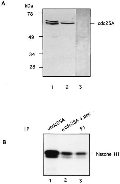
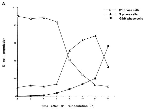
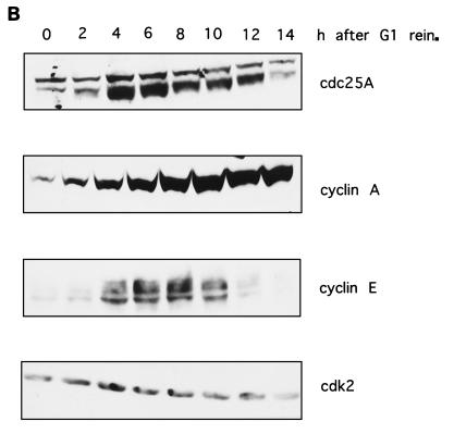
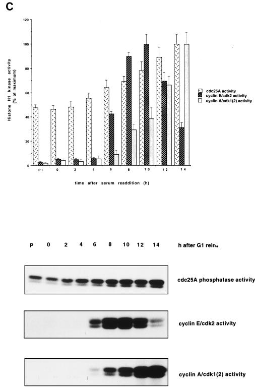
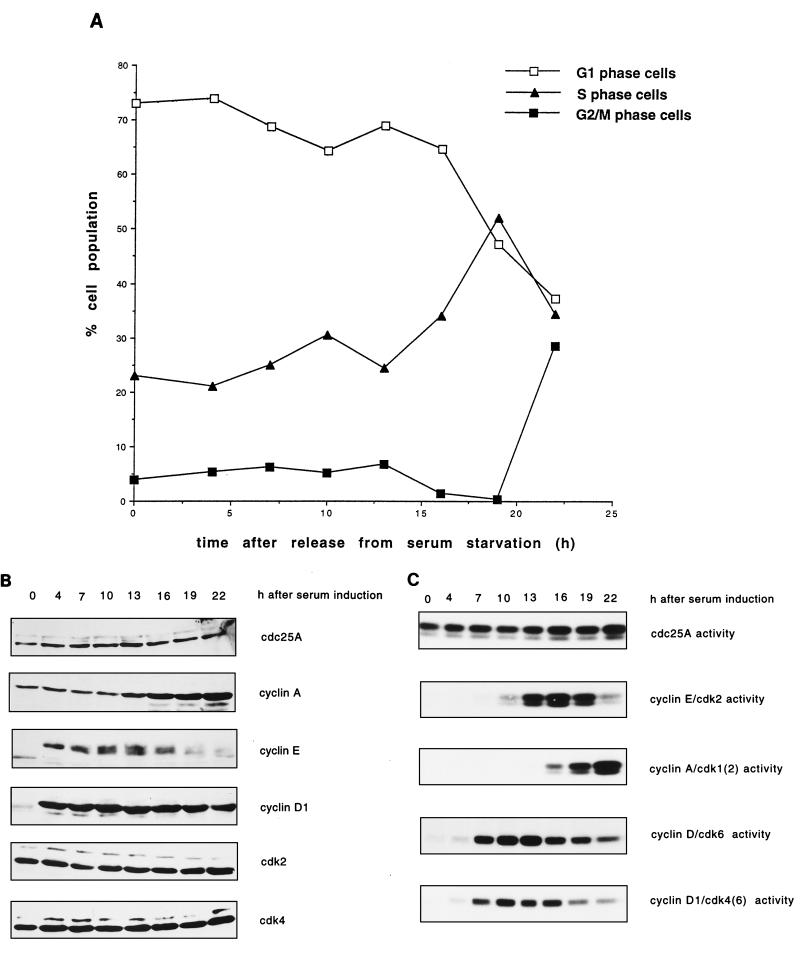
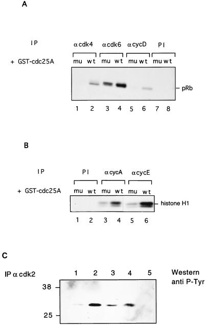
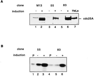
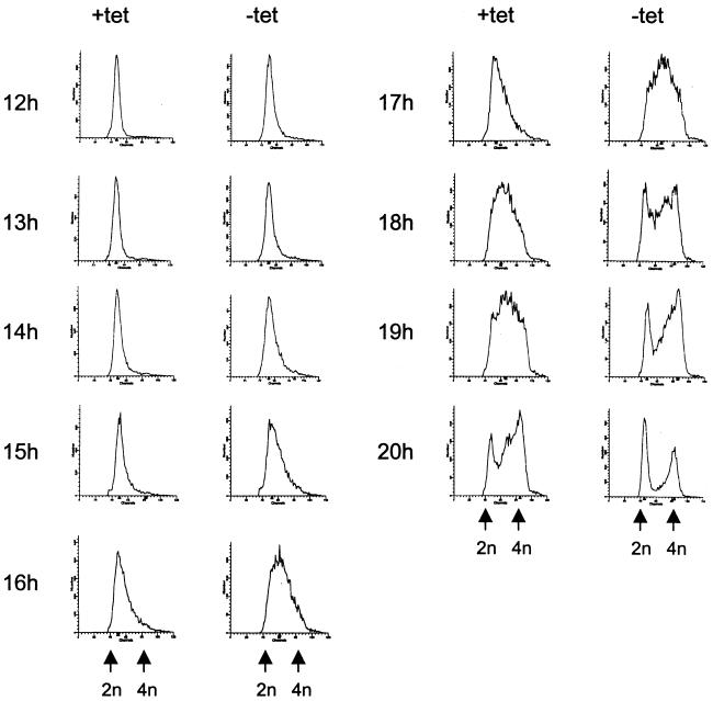
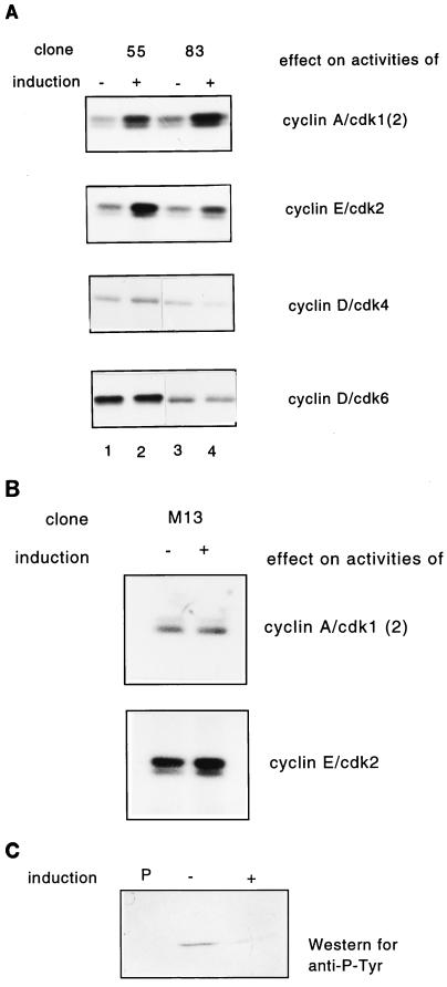
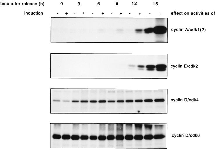
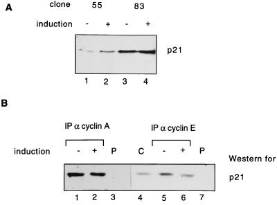
Human Cdc25 phosphatases play important roles in cell cycle regulation by removing inhibitory phosphates from tyrosine and threonine residues of cyclin-dependent kinases. Three human Cdc25 isoforms, A, B, and C, have been discovered. Cdc25B and Cdc25C play crucial roles at the G(2)/M transition. In the present study, we have investigated the function of human Cdc25A phosphatase. Cell lines that express human Cdc25A in an inducible manner have been generated. Ectopic expression of Cdc25A accelerates the G(1)/S-phase transition, indicating that Cdc25A controls an event(s) that is rate limiting for entry into S phase. Furthermore, we carried out a detailed analysis of the expression and activation of human Cdc25A. Activation of endogenous Cdc25A occurs during late G(1) phase and increases in S and G(2) phases. We further demonstrate that Cdc25A is activated at the same time as cyclin E- and cyclin A-dependent kinases. In vitro, Cdc25A dephosphorylates and activates the cyclin-Cdk complexes that are active during G(1). Overexpression of Cdc25A in the inducible system, however, leads to a premature activation of both cyclin E-Cdk2 and cyclin A-Cdk2 complexes, while no effect of cyclin D-dependent kinases is observed. Furthermore, Cdc25A overexpression induces a tyrosine dephosphorylation of Cdk2. These results suggest that Cdc25A is an important regulator of the G(1)/S-phase transition and that cyclin E- and cyclin A-dependent kinases act as direct targets.


Figures

FIG. 1
FIG. 1
Characterization of Cdc25A antibodies. (A) Extracts of HeLa cells (lanes 1 and 3) and HS68 cells (lane 2) were resolved by SDS-PAGE, transferred to nitrocellulose, and probed with affinity-purified Cdc25A antibodies raised against the full-length protein (lanes 1 and 2) or preimmune serum (lane 3). (B) Cdc25A was immunoprecipitated from HeLa cell extracts with a C-terminal peptide antibody in the absence (lane 1) or presence (lane 2) of antigenic peptide and incubated with tyrosine-phosphorylated inactive cyclin B-Cdk1 complex. Activation of cyclin B-Cdk1 caused by Cdc25A was monitored on histone H1 as substrate. Lane 3 shows a control immunoprecipitation using preimmune (PI) serum. IP, immunoprecipitant.
FIG. 2
FIG. 2
Protein levels and activity of Cdc25A and G1 cyclins-Cdks in HeLa cells. (A) Cell cycle distribution. HeLa cells grown in suspension were separated on the basis of size by centrifugal elutriation. G1-enriched cells were reinoculated in fresh medium and collected at the indicated time intervals. Cells were stained with propidium iodide and analyzed by flow cytometry. The graph shows the percentages of cells in G1, S, and G2/M phases. (B) Immunoblot analysis. Total cell extracts from the elutriated samples of panel A were analyzed by SDS-PAGE and immunoblotted with specific antibodies against Cdc25A, cyclins E and A, and Cdk2. (C) Cdc25A phosphatase activity and cyclin E- and cyclin A-associated kinase activities. Extracts were prepared at the indicated times from the elutriated samples, immunoprecipitated with Cdc25A antiserum, and incubated with tyrosine-phosphorylated inactive cyclin B-Cdk1 complex. Activation of cyclin B-Cdk1, caused by Cdc25A, was monitored on histone H1 as substrate. Cyclin E- and cyclin A-associated kinase activity was tested on histone H1 as substrate. P denotes control immunoprecipitations with a preimmune serum. The results are the means of two independent experiments. rein., reinoculation.
FIG. 2
FIG. 2
Protein levels and activity of Cdc25A and G1 cyclins-Cdks in HeLa cells. (A) Cell cycle distribution. HeLa cells grown in suspension were separated on the basis of size by centrifugal elutriation. G1-enriched cells were reinoculated in fresh medium and collected at the indicated time intervals. Cells were stained with propidium iodide and analyzed by flow cytometry. The graph shows the percentages of cells in G1, S, and G2/M phases. (B) Immunoblot analysis. Total cell extracts from the elutriated samples of panel A were analyzed by SDS-PAGE and immunoblotted with specific antibodies against Cdc25A, cyclins E and A, and Cdk2. (C) Cdc25A phosphatase activity and cyclin E- and cyclin A-associated kinase activities. Extracts were prepared at the indicated times from the elutriated samples, immunoprecipitated with Cdc25A antiserum, and incubated with tyrosine-phosphorylated inactive cyclin B-Cdk1 complex. Activation of cyclin B-Cdk1, caused by Cdc25A, was monitored on histone H1 as substrate. Cyclin E- and cyclin A-associated kinase activity was tested on histone H1 as substrate. P denotes control immunoprecipitations with a preimmune serum. The results are the means of two independent experiments. rein., reinoculation.
FIG. 3
FIG. 3
Protein levels and activities of Cdc25A and G1 cyclins-Cdks in Hs68 human fibroblasts. (A) Hs68 cells were synchronized by serum starvation and released by addition of DMEM containing 20% FCS. Samples were taken for flow cytometry at the indicated times. (B) Total cell extracts were analyzed by SDS-PAGE and immunoblotted with Cdc25A-specific antibodies or with antibodies against the different G1 Cdks and cyclins. (C) Phosphatase and kinase activities from the same time points were determined as described for Fig. 2C. The activities of cyclin D immunocomplexes were measured as phosphorylation of pRb.
FIG. 4
FIG. 4
Dephosphorylation and activation of cyclin-Cdk complexes by Cdc25A in vitro. (A) Cyclin D-Cdk4 or cyclin D-Cdk6 complexes were immunoprecipitated from exponentially growing IMR-90 cells by using antibodies against Cdk4 (lanes 1 and 2), Cdk6 (lanes 3 and 4), and cyclin D1 (lanes 5 and 6) or preimmune sera (lanes 7 and 8) and incubated with GST-Cdc25A (lanes 2, 4, 6, and 8) or GST-Cdc25A (C430S) (lanes 1, 3, 5, and 7). Activities of the complexes were determined on pRb as substrate. (B) Cyclin E-Cdk2 and cyclin A-Cdk2 complexes were immunoprecipitated from exponentially growing HeLa cells by using antibodies against cyclin A (lanes 3 and 4) and cyclin E (lanes 5 and 6) or preimmune serum (lanes 1 and 2) and incubated with GST-Cdc25A (lanes 2, 4, and 6) or GST-Cdc25A C430S (lanes 1, 3, and 5). Kinase activities were measured on histone H1 as substrate. (C) Cdk2 was immunoprecipitated from exponential HeLa cell extract and incubated with either GST-Cdc25A (lane 1), GST-Cdc25A (C430S) (lane 2), GST-Cdc25A in the presence of 1 mM sodium orthovanadate (lane 3), or buffer alone (lane 4); blotted; and incubated with antiphosphotyrosine antibodies. In lane 5, the immunoprecipitation was carried out with preimmune serum. Numbers at left indicate molecular masses in kilodaltons. IP, immunoprecipitant; PI, preimmune serum; mu, mutant; wt, wild type.
FIG. 5
FIG. 5
Inducible expression and activities of human Cdc25A in R12 cells. Clones M13 (lanes 1 and 2), 55 (lanes 3 and 4), and 83 (lanes 5 and 6) were grown in the presence (no induction of Cdc25A) and absence (induction of Cdc25A) of tetracycline for 48 h. Cells were then lysed as described in Materials and Methods. (A) Immunoblot analysis. Protein extracts from induced (+) and noninduced (−) cells were run on an SDS–10% polyacrylamide gel and blotted on nitrocellulose. The blot was then incubated with Cdc25A antibodies. HeLa cell lysates (lane 7) were used as controls. (B) Protein (1 mg) was used to analyze Cdc25A phosphatase activity by using inactive tyrosine-phosphorylated cyclin B-Cdk1 as substrate. Cyclin B-Cdk1 activation was determined by histone H1 phosphorylation. Lanes 1 and 4, control immunoprecipitations with preimmune serum; lanes 2 and 5, activity without induction of Cdc25A; lanes 3 and 6, activity with induction of Cdc25A.
FIG. 6
FIG. 6
Effects of Cdc25A overexpression on cell cycle progression of rat-1 cells. Clone 55 was arrested in G0 with DMEM containing 0.1% FCS in the presence (+) (no induction of Cdc25A) or absence (−) (induction of Cdc25A) of tetracycline (tet) for 48 h. The cells were released with 10% FCS (with or without tetracycline). Cells were harvested at the indicated times after release. (A) The samples were fixed, stained with propidium iodide, and analyzed by flow cytometry. (B) One hour before harvest, 30 μM BrdU was added. Cells were double labelled with anti-BrdU antibodies and propidium iodide. BrdU staining of the cells was analyzed by flow cytometry. The figure indicates the percentage of S-phase (BrdU)-labelled cells.
FIG. 6
FIG. 6
Effects of Cdc25A overexpression on cell cycle progression of rat-1 cells. Clone 55 was arrested in G0 with DMEM containing 0.1% FCS in the presence (+) (no induction of Cdc25A) or absence (−) (induction of Cdc25A) of tetracycline (tet) for 48 h. The cells were released with 10% FCS (with or without tetracycline). Cells were harvested at the indicated times after release. (A) The samples were fixed, stained with propidium iodide, and analyzed by flow cytometry. (B) One hour before harvest, 30 μM BrdU was added. Cells were double labelled with anti-BrdU antibodies and propidium iodide. BrdU staining of the cells was analyzed by flow cytometry. The figure indicates the percentage of S-phase (BrdU)-labelled cells.
FIG. 7
FIG. 7
Effect of Cdc25A overproduction on cyclin-dependent kinase activities in asynchronously growing HeLa cells. (A) Wild-type Cdc25A was not induced (lanes 1 and 3) or induced (lanes 2 and 4) by tetracycline addition or removal, respectively, for 24 h. The histone H1 kinase activities associated with cyclin A and cyclin E or pRb kinase activities associated with Cdk4 and Cdk6 immunoprecipitates were analyzed. The experiment was performed with clones 55 (lanes 1 and 2) and 83 (lanes 3 and 4). (B) Mutant Cdc25A was either noninduced (−) or induced (+) as described for panel A, and the histone H1 kinase activities associated with cyclin A and cyclin E were analyzed. (C) Wild-type Cdc25A (clone 83) was either noninduced (−) or induced (+) as described for panel A. Cdk2 was immunoprecipitated and analyzed by immunoblotting with antiphosphotyrosine antibodies. P denotes a control precipitation with preimmune serum.
FIG. 8
FIG. 8
Effect of Cdc25A overproduction on the activities of G1 Cdks-cyclins in synchronized R12 cells. Cells were synchronized and induced as described in the legend to Fig. 6. Extracts were prepared after the times indicated and used to determine G1 Cdk-cyclin activities. The kinases were immunoprecipitated with cyclin A, cyclin E, Cdk4, and Cdk6 antibodies. Cyclin A- and cyclin E-associated kinase activities were measured on histone H1 as substrate, and Cdk4 and Cdk6 activities were measured on pRb as substrate.
FIG. 9
FIG. 9
Expression and cyclin binding of p21 in Cdc25A-overproducing cells. (A) Clones 55 and 83 were induced (+) or noninduced (−) by tetracycline removal and addition for 24 h. Total levels of p21 after immunoblotting with p21 antibodies without induction (lanes 1 and 3) and with induction of Cdc25A (lanes 2 and 4) are shown. (B) Clone 83 was induced as described above. Amounts of p21 bound to immunoprecipitated cyclin A-Cdk2 (lanes 1 and 2) or cyclin E-Cdk2 (lanes 5 and 6) complexes or to control immunoprecipitations (with preimmune [P] serum) (lanes 3 and 7) were determined by immunoblotting with p21 antibodies. Lane 4 shows the migration and levels of p21 in whole rat-1 cell (C) lysates. IP, immunoprecipitant.

References

    1. Cai G, Dynlacht B. Activity and nature of p21 WAF1 complexes during the cell cycle. Proc Natl Acad Sci USA. 1998;95:12254–12259. - PMC - PubMed
    1. Coqueret O, Bérubé G, Nepveu A. The mammalian Cut homeodomain protein functions as a cell-cycle-dependent transcriptional repressor which downmodulates p21WAF1/CIP/SDI1 in S phase. EMBO J. 1998;17:4680–4694. - PMC - PubMed
    1. Draetta G, Beach D. Activation of cdc2 protein kinase during mitosis in human cells: cell cycle-dependent phosphorylation and subunit rearrangement. Cell. 1988;54:17–26. - PubMed
    1. Fauman E, Cogswell J, Lovejoy B, Rocque W, Holmes W, Montana V, Piwnica-Worms H, Rink M, Saper M. Crystal structure of the catalytic domain of the human cell cycle control phosphatase, Cdc25A. Cell. 1998;93:617–625. - PubMed
    1. Gabrielli B, De Souza C, Tonks I, Clark J, Hayward N, Ellem K. Cytoplasmic accumulation of Cdc25B phosphatase in mitosis triggers centrosomal microtubule nucleation in HeLa cells. J Cell Sci. 1996;109:1081–1093. - PubMed

Publication types