Restrictive pericarditis
- PMID: 10455096
- PMCID: PMC1729164
- DOI: 10.1136/hrt.82.3.389
Restrictive pericarditis
Abstract
Background: Pericardial thickening is an uncommon complication of cardiac surgery.
Objectives: To study pericardial thickening as the cause of severe postoperative venous congestion.
Subjects: Two men, one with severe aortic stenosis and single coronary artery disease, and one with coronary artery disease after an old inferior infarction. Both had coronary artery bypass grafting surgery.
Methods: Magnetic resonance imaging (MRI), Doppler echocardiography, and cardiac catheterisation.
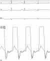
Results: Venous pressure was raised in both patients. MRI showed mildly thickened pericardium, and cardiac catheterisation indicated diastolic equalization of pressures in the four chambers. Jugular venous pulse showed a dominant "Y" descent coinciding with early diastolic flow in the superior vena cava, and mitral and tricuspid Doppler forward flow proved restrictive physiology. The clinical background suggested pericardial disease so both patients had pericardiectomy. This proved the pericardium to be thickened; the extent of fibrosis also involved the epicardium.
Conclusions: Although rare, restrictive pericarditis (restrictive ventricular physiology resulting from pericardial disease) should be considered to be a separate diagnostic entity because its pathological basis and treatment are different from intrinsic myocardial disease. This diagnosis may be confirmed by standard investigational techniques or may require diagnostic thoracotomy.
Figures


Publication types
MeSH terms
LinkOut - more resources
Full Text Sources
Medical
