

Regulation of epidermal homeostasis through P2Y2 receptors
- PMID: 10455326
- PMCID: PMC1566136
- DOI: 10.1038/sj.bjp.0702653
Regulation of epidermal homeostasis through P2Y2 receptors
Abstract
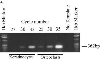
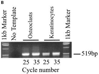
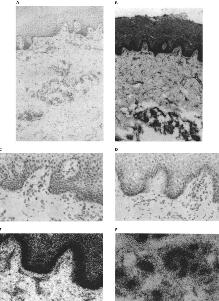
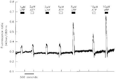
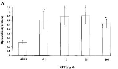
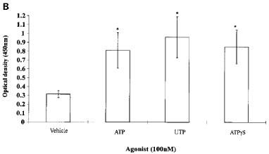
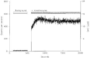
1. Previous studies have indicated a role for extracellular ATP in the regulation of epidermal homeostasis. Here we have investigated the expression of P2Y2 receptors by human keratinocytes, the cells which comprise the epidermis. 2. Reverse transcriptase-polymerase chain reaction (RT - PCR) revealed expression of mRNA for the G-protein-coupled, P2Y2 receptor in primary cultured human keratinocytes. 3. In situ hybridization studies of skin sections revealed that P2Y2 receptor transcripts were expressed in the native tissue. These studies demonstrated a striking pattern of localization of P2Y2 receptor transcripts to the basal layer of the epidermis, the site of cell proliferation. 4. Increases in intracellular free Ca2+ concentration ([Ca2+]i) in keratinocytes stimulated with ATP or UTP demonstrated the presence of functional P2Y receptors. 5. In proliferation studies based on the incorporation of bromodeoxyuridine (BrdU), ATP, UTP and ATPgammaS were found to stimulate the proliferation of keratinocytes. 6. Using a real-time firefly luciferase and luciferin assay we have shown that under static conditions cultured human keratinocytes release ATP. 7. These findings indicate that P2Y2 receptors play a major role in epidermal homeostasis, and may provide novel targets for therapy of proliferative disorders of the epidermis, including psoriasis.
Figures
Similar articles
-
Human keratinocytes express multiple P2Y-receptors: evidence for functional P2Y1, P2Y2, and P2Y4 receptors.J Invest Dermatol. 2003 Mar;120(3):440-7. doi: 10.1046/j.1523-1747.2003.12050.x. J Invest Dermatol. 2003. PMID: 12603858
-
P2Y2 receptor up-regulation induced by guanosine or UTP in rat brain cultured astrocytes.Int J Immunopathol Pharmacol. 2006 Apr-Jun;19(2):293-308. doi: 10.1177/039463200601900207. Int J Immunopathol Pharmacol. 2006. PMID: 16831297
-
P2Y2 receptors are expressed by human osteoclasts of giant cell tumor but do not mediate ATP-induced bone resorption.Bone. 1998 Mar;22(3):195-200. doi: 10.1016/s8756-3282(97)00280-9. Bone. 1998. PMID: 9514211
-
Molecular determinants of P2Y2 nucleotide receptor function: implications for proliferative and inflammatory pathways in astrocytes.Mol Neurobiol. 2005;31(1-3):169-83. doi: 10.1385/MN:31:1-3:169. Mol Neurobiol. 2005. PMID: 15953819 Review.
-
P2Y receptors in the nervous system: molecular studies of a P2Y2 receptor subtype from NG108-15 neuroblastoma x glioma hybrid cells.Prog Brain Res. 1999;120:33-43. doi: 10.1016/s0079-6123(08)63544-x. Prog Brain Res. 1999. PMID: 10550986 Review. No abstract available.
Cited by
-
Purine- and pyrimidine-induced responses and P2Y receptor characterization in the hamster proximal urethra.Br J Pharmacol. 2005 Feb;144(4):510-8. doi: 10.1038/sj.bjp.0706047. Br J Pharmacol. 2005. PMID: 15655529 Free PMC article.
-
Chemosensory information processing between keratinocytes and trigeminal neurons.J Biol Chem. 2014 Jun 20;289(25):17529-40. doi: 10.1074/jbc.M113.499699. Epub 2014 May 1. J Biol Chem. 2014. PMID: 24790106 Free PMC article.
-
P2Y2 nucleotide receptors mediate metalloprotease-dependent phosphorylation of epidermal growth factor receptor and ErbB3 in human salivary gland cells.J Biol Chem. 2010 Mar 5;285(10):7545-55. doi: 10.1074/jbc.M109.078170. Epub 2010 Jan 11. J Biol Chem. 2010. PMID: 20064929 Free PMC article.
-
Purinergic receptors play a key role in shock wave-induced proliferation.Sci Rep. 2025 May 31;15(1):19138. doi: 10.1038/s41598-025-02955-3. Sci Rep. 2025. PMID: 40450090 Free PMC article.
-
Melittin modulates keratinocyte function through P2 receptor-dependent ADAM activation.J Biol Chem. 2012 Jul 6;287(28):23678-89. doi: 10.1074/jbc.M112.362756. Epub 2012 May 21. J Biol Chem. 2012. PMID: 22613720 Free PMC article.
References
-
- BARRANDON Y., GREEN H. Cell migration is essential for sustained growth of keratinocyte colonies: the roles of transforming growth factor-α and epidermal growth factor. Cell. 1984;50:1131–1137. - PubMed
-
- BOARDER M.R., HOURANI S.M.O. The regulation of vascular function by P2 receptors: multiple sites and multiple receptors. Trends Pharmacol. Sci. 1998;19:99–107. - PubMed
-
- BODIN P., BURNSTOCK G. ATP-stimulated release of ATP by human endothelial cells. J. Cardiovasc. Pharmacol. 1996;27:872–875. - PubMed
-
- BOWLER W.B., BIRCH M.A., GALLAGHER J.A., BILBE G. Identification and cloning of human P2u purinoceptor present in osteoclastoma, bone, and osteoblasts. J. Bone Min. Res. 1995;10:1137–1145. - PubMed
Publication types
MeSH terms
Substances
Grants and funding
LinkOut - more resources
Full Text Sources
Other Literature Sources
Molecular Biology Databases
Miscellaneous
