

Characterization of the interaction between Yersinia enterocolitica biotype 1A and phagocytes and epithelial cells in vitro
- PMID: 10456876
- PMCID: PMC96754
- DOI: 10.1128/IAI.67.9.4367-4375.1999
Characterization of the interaction between Yersinia enterocolitica biotype 1A and phagocytes and epithelial cells in vitro
Abstract
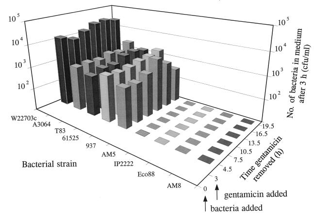
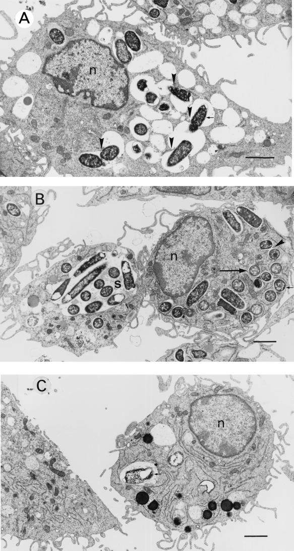
Yersinia enterocolitica strains of biotype 1A are increasingly being recognized as etiological agents of gastroenteritis. However, the mechanisms by which these bacteria cause disease differ from those of highly invasive, virulence plasmid-bearing Y. enterocolitica strains and are poorly understood. We have investigated several biotype 1A strains of diverse origin for their ability to resist killing by professional phagocytes. All strains were rapidly killed by polymorphonuclear leukocytes but persisted within macrophages (activated with gamma interferon) to a significantly greater extent (survival = 40.5% +/- 17.4%) than did Escherichia coli HB101 (9.3% +/- 0.7%; P = 0.0001). Strains isolated from symptomatic patients were significantly more resistant to killing by macrophages (survival = 48.9% +/- 19.5%) than were strains obtained from food or the environment (survival = 32.1% +/- 10.3%; P = 0.04). Some strains which had been ingested by macrophages or HEp-2 epithelial cells showed a tendency to reemerge into the tissue culture medium over a period lasting several hours. This phenomenon, which we termed "escape," was observed in 14 of 15 strains of clinical origin but in only 3 of 12 nonclinical isolates (P = 0.001). The capacity of bacteria to escape from cells was not directly related to their invasive ability. To determine if escape was due to host cell lysis, we used a variety of techniques, including lactate dehydrogenase release, trypan blue exclusion, and examination of infected cells by light and electron microscopy, to measure cell viability and lysis. These studies established that biotype 1A Y. enterocolitica strains were able to escape from macrophages or epithelial cells without causing detectable cytolysis, suggesting that escape was achieved by a process resembling exocytosis. The observations that biotype 1A Y. enterocolitica strains of clinical origin are significantly more resistant to killing by macrophages and significantly more likely to escape from host cells than are strains of nonclinical origin suggest that these properties may account for the virulence of these bacteria.
Figures
References
-
- Apicella M A, Ketterer M, Lee F K N, Zhou D, Rice P A, Blake M S. The pathogenesis of gonococcal urethritis in men: confocal and immunoelectron microscopic analysis of urethral exudates from men infected with Neisseria gonorrhoeae. J Infect Dis. 1996;173:636–646. - PubMed
Publication types
MeSH terms
LinkOut - more resources
Full Text Sources
