. 1999 Sep;67(9):4456-62.
doi: 10.1128/IAI.67.9.4456-4462.1999.

Role of Mycoplasma penetrans endonuclease P40 as a potential pathogenic determinant

Affiliations

Role of Mycoplasma penetrans endonuclease P40 as a potential pathogenic determinant

M Bendjennat et al. Infect Immun. 1999 Sep.

Abstract

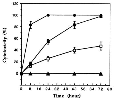
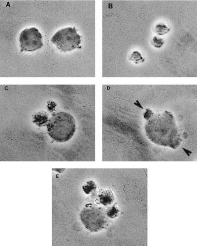
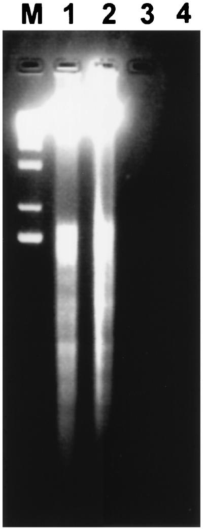
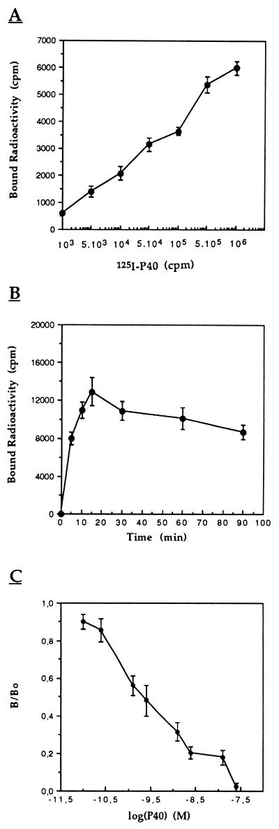
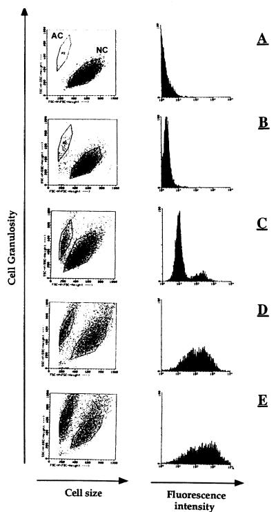
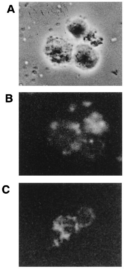
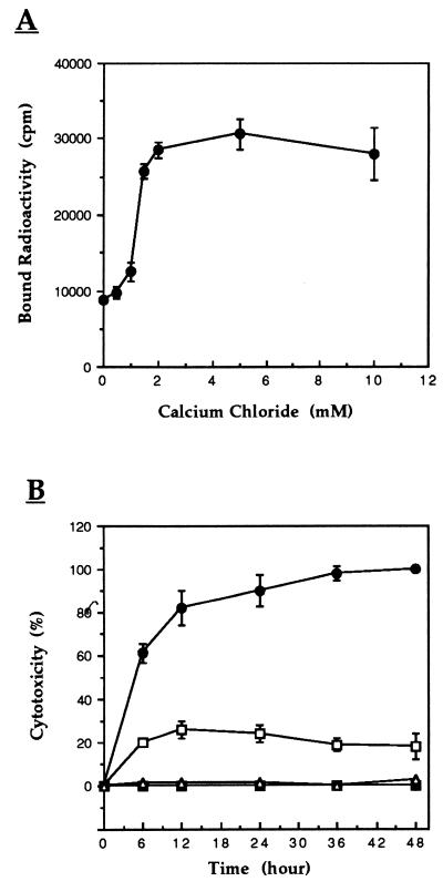
Recently, we reported the purification to homogeneity and characterization of Ca(2+)- and Mg(2+)-dependent endonuclease P40 produced by Mycoplasma penetrans (M. Bendjennat, A. Blanchard, M. Loutfi, L. Montagnier, and E. Bahraoui, J. Bacteriol. 179; 2210-2220, 1997), a mycoplasma which was isolated for the first time from the urine of human immunodeficiency virus-infected patients. To evaluate how this nuclease could interact with host cells, we tested its effect on CEM and Molt-4 lymphocytic cell lines and on peripheral blood mononuclear cells. We observed that 10(-7) to 10(-9) M P40 is able to mediate a cytotoxic effect. We found that 100% of cells were killed after 24 h of incubation with 10(-7) M P40 while only 40% cytotoxicity was obtained after 72 h of incubation with 10(-9) M P40. Phase-contrast microscopy observations of P40-treated cells revealed morphological changes, including pronounced blebbing of the plasma membrane and cytoplasmic shrinkage characteristic of programmed cell death, which is in agreement with the internucleosomal fragmentation of P40-treated cell DNA as shown by agarose gel electrophoresis. We showed that (125)I-radiolabeled or fluorescein isothiocyanate-labeled P40 was able to bind specifically in a dose-dependent manner to the cell membrane of CEM cells, which suggested that the cytotoxicity of P40 endonuclease was mediated by its interaction with the cell surface receptor(s). The concentration of unlabeled P40 required to inhibit by 50% the formation of (125)I-P40-CEM complexes was about 3 x 10(-9) M, indicating a high-affinity interaction. Both P40 interaction and cytotoxicity are Ca(2+) dependent. Our results suggest that the cytotoxicity of M. penetrans observed in vitro is mediated at least partially by secreted P40, which, after interaction with host cells, can induce an apoptosis-like death. These results strongly suggest a major role of mycoplasmal nucleases as potential pathogenic determinants.


Figures

FIG. 1
FIG. 1
Cytotoxicity of M. penetrans endonuclease P40 toward CEM cells. CEM cells were washed and seeded in 96-well plates (104 cells/200 μl of culture medium) and exposed at time zero to endonuclease P40 (0 M [▴], 10−9 M [□], 10−8 M [■], or 10−7 M [●]) at 37°C. Percent cytotoxicity was determined at the defined time points and is calculated as the difference between control (100%) and percent survival. Values are means ± standard errors from triplicate experiments.
FIG. 2
FIG. 2
The effects of P40 protein on CEM cells in vitro were assessed by phase-contrast microscopy. CEM cells (104) were cultured in flat-bottom microtiter plates in the absence or presence of 10−8 M P40. After 24 h, photomicrographs were taken. (A) Negative control; (B to E) morphological changes of cells incubated in the presence of P40. Arrows indicate apoptosis-like bodies.
FIG. 3
FIG. 3
Endonuclease P40-induced internucleosomal DNA fragmentation. Small supernatant DNA after centrifugation of detergent-lyzed cells was separated on a 2% agarose gel. CEM cells (lane 1) and PBMCs (lane 2) (106) were incubated with P40 (10−8 M) for 24 h at 37°C in culture medium. Lanes 3 and 4 contain CEM and PBMC negative controls, respectively. Lane M contains the 1-kb DNA ladder used as a standard.
FIG. 4
FIG. 4
Direct radiolabeling assay analysis of 125I-P40 binding to CEM cells. (A) A 50-μl volume of cells (106) was incubated with 50 μl of various 125I-P40 dilutions (0 to 106 cpm; 0 to 10−9 M). (B) Time course of 125I-P40 (106 cpm; 10−9 M) binding to CEM cells (8 × 10⁶). (C) Inhibition of 125I-P40 binding to CEM cells (106) by native protein at 10−8 M and 37°C. B/B0 corresponds to the binding obtained in the presence of competitor/binding obtained in the absence of competitor. After the cells were washed twice with PBS–BSA buffer, bound radioactivity was counted. Values are means ± standard errors of triplicate experiments.
FIG. 5
FIG. 5
Direct fluorescence labeling of P40 bound to CEM cells and inducing a cytopathic effect. A total of 106 cells were incubated with different dilutions of FITC-conjugated P40 (negative control [A] and 1 × 10⁻⁹ [B], 1 × 10⁻⁸ [C], 5 × 10⁻⁷ [D], and 1 × 10⁻⁷ M [E], respectively) for 4 h at 37°C, in 100 μl of PBS, BSA buffer. After the cells were washed, they were screened by flow cytometry analysis. NC and AC, normal cells and altered cells, respectively.
FIG. 6
FIG. 6
Distribution of staining on fluorescence analysis of CEM cells from Fig. 5E by fluorescence microscopy. Phase-contrast (A) and fluorescence (B) microscopy observations. (C) Another field of fluorescent cells.
FIG. 7
FIG. 7
(A) Effect of Ca2+ on the avidity of 125I-P40 binding. A 50-μl volume of 125I-P40 (2 × 10⁶ cpm, 2 × 10⁻⁹ M) in PBS-BSA buffer containing several dilutions of CaCl2 was added to 50 μl of CEM cells (106) and incubated at 37°C for 15 min. After the cells were washed twice with buffer, bound radioactivity was counted. The effect of Ca2+ on 125I-P40 binding activity was abolished in the presence of EDTA and was reproducible in Tris and morpholineproponesulfonic acid (MOPS) buffer instead of PBS. (B) Effect of Ca2+ on cytotoxicity induced by endonuclease P40. The cytopathic effect of P40 was assessed as described in the legend to Fig. 1 (104 cells, 10−8 M P40), except that cells were cultivated in calcium-free medium. Percent cytotoxicity was determined at the defined time points. ■ and ▵, cells cultivated with and without exogenous CaCl2, respectively; □, P40-treated cells; ●, P40-treated cells in the presence of CaCl2. Data are the means ± standard errors.

Similar articles

Cited by

References

    1. Alnemri E S, Litwack G. Glucocorticoid-induced lymphocytolysis is not mediated by an induced endonuclease. J Biol Chem. 1989;264:4104–4111. - PubMed
    1. Andreev J, Borovsky Z, Rosenshine I, Rottem S. Invasion of HeLa cells by Mycoplasma penetrans and the induction of tyrosine phosphorylation of a 145 kDa host cell protein. FEMS Microbiol Lett. 1995;132:189–194. - PubMed
    1. Bendjennat M, Blanchard A, Loutfi M, Montagnier L, Bahraoui E. Purification and characterization of Mycoplasma penetrans Ca2+/Mg2+-dependent endonuclease. J Bacteriol. 1997;179:2210–2220. - PMC - PubMed
    1. Benedik M J, Strych U. Serratia marcescens and its extracellular nuclease. FEMS Microbiol Lett. 1998;165:1–13. - PubMed
    1. Blanchard A, Montagnier L. Aids-associated mycoplasmas. Annu Rev Microbiol. 1994;48:687–712. - PubMed

Publication types

LinkOut - more resources