. 1999 Sep;67(9):4477-84.
doi: 10.1128/IAI.67.9.4477-4484.1999.

Interaction of Leishmania gp63 with cellular receptors for fibronectin

Affiliations

Interaction of Leishmania gp63 with cellular receptors for fibronectin

A Brittingham et al. Infect Immun. 1999 Sep.

Abstract

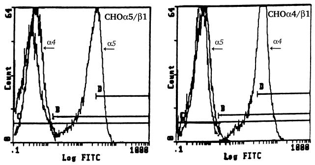
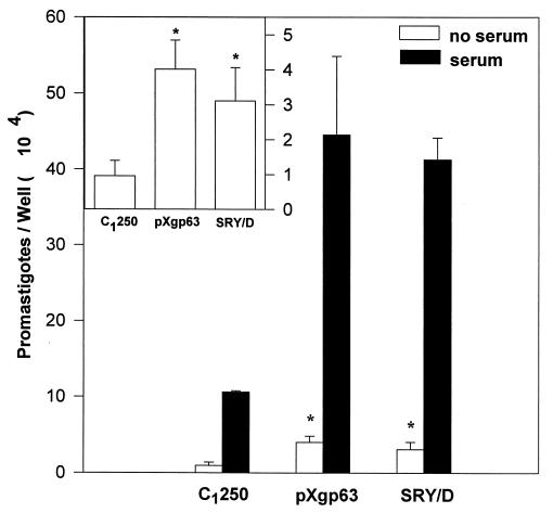
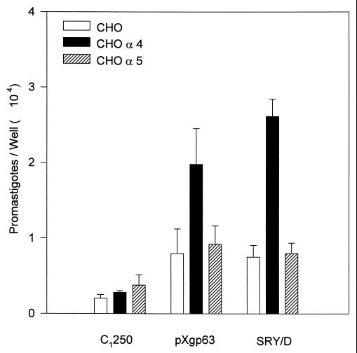
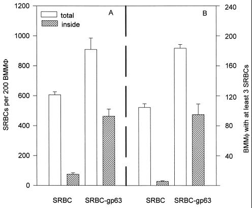
The most abundant protein on the surface of the promastigote form of the protozoan parasites Leishmania spp. is a 63-kDa molecule, designated gp63 or leishmanolysin. Because gp63 has been shown to possess fibronectin-like properties, we examined the interaction of gp63 with the cellular receptors for fibronectin. We measured the direct binding of Leishmania to human macrophages or to transfected mammalian cells expressing human fibronectin receptors. Leishmania expressing gp63 exhibited modest but reproducible adhesion to human macrophages and to transfected CHO cells expressing alpha4/beta1 fibronectin receptors. In both cases, this interaction depended on gp63 but occurred independently of the SRYD sequence of gp63, because parasites expressing gp63 with a mutated SRYD sequence bound to macrophages and alpha4/beta1 receptor-expressing cells as well as did wild-type parasites. The contribution of gp63 to parasite adhesion was more pronounced when the assays were performed in the presence of complement, suggesting that the receptors for complement and fibronectin may cooperate to mediate the efficient adhesion of parasites to macrophages. The interaction of gp63 with fibronectin receptors may also play an important role in parasite internalization by macrophages. Erythrocytes to which gp63 was cross-linked were efficiently phagocytized by macrophages, whereas control erythrocytes opsonized with complement alone bound to macrophages but remained peripherally attached to the outside of the cell. Similarly, parasites expressing wild-type gp63 were rapidly and efficiently phagocytized by resting macrophages, whereas parasites lacking gp63 were internalized more slowly. This rapid internalization of gp63-expressing parasites was dependent on the beta1 integrins, because pretreatment of macrophages with monoclonal antibodies to the beta1 integrins decreased the internalization of gp63-expressing parasites. These observations indicate that complement receptors are the primary mediators of parasite adhesion; however, maximal parasite adhesion and internalization may require the participation of the beta1 integrins, which recognize fibronectin-like molecules such as gp63 on the surface of the parasite.


Figures

FIG. 1
FIG. 1
Cell surface expression of β1 integrins on transfected CHO-B2 cells. CHO-B2/α5 cells (left panel) or CHO-B2/α4 cells (right panel) were incubated individually with MAb to detect either α4 (HP2/1) or α5 (SAM1) receptor expression on their surface. Cells were stained with FITC-conjugated goat anti-mouse immunoglobulin G (Jackson ImmunoResearch) and analyzed by flow cytometry. Profiles of cells stained with each antibody were overlaid and compared to those of cells stained with secondary antibody alone.
FIG. 2
FIG. 2
Binding of radiolabeled L. amazonensis gp63 variants to human monocyte-derived macrophages. Radiolabeled promastigotes were added to monolayers of human monocyte-derived macrophages at a 10:1 ratio in the presence (filled bars) or absence (open bars) of 4% C8D serum as a source of opsonic complement. Following 45 min of incubation and extensive washing, the number of promastigotes associated with human monocyte-derived macrophages was determined as described in Materials and Methods. Values represent the mean of four experiments, each performed in triplicate, ± the standard error. Statistical significance at the 95% confidence level (P, ≤0.05), determined with the Student’s t test, is denoted by an asterisk. The inset is an enlargement of the binding of promastigotes to macrophages in the absence of serum.
FIG. 3
FIG. 3
Binding of L. amazonensis gp63 variants to CHO cells expressing human fibronectin receptors. Radiolabeled promastigotes were added to monolayers of CHO cells at a 50:1 ratio for 1 h at 37°C. The number of promastigotes associated with fibronectin receptor-deficient CHO cells (open bars) and CHO cells transfected with cDNA for the human α4 receptor (CHO α4) (solid bars) or the human α5 receptor (CHO α5) (hatched bars) was determined as described in Materials and Methods. Determinations were performed in triplicate, and values are expressed as the mean ± standard deviation. This experiment is representative of three experiments.
FIG. 4
FIG. 4
Binding of L. amazonensis gp63 variants to transfected CHO cells expressing human complement receptors. Radiolabeled promastigotes were added to monolayers of transfected CHO cells expressing Mac-1 (open bars) or CR1 (hatched bars) or control transfected cells (F185.1) expressing a truncated form of intracellular adhesion molecule 1 (solid bars), at a 50:1 ratio, in either the presence (+S) or the absence (−S) of serum. The number of promastigotes associated with each monolayer is expressed as the mean ± standard deviation of triplicate determinations. This experiment is representative of three experiments.
FIG. 5
FIG. 5
Binding and internalization of SRBC-gp63 by murine bone marrow-derived macrophages (BMMφ). SRBC or SRBC-gp63 were added to parallel monolayers of murine bone marrow-derived macrophages. After 1 h, all monolayers were washed to remove nonadherent cells. To determine the total number of erythrocytes associated with 200 macrophages (total), washed monolayers were fixed in 2.5% glutaraldehyde and processed for light microscopy (open bars). To determine the number of erythrocytes inside macrophages (inside), monolayers were rinsed for 15 s in water to lyse extracellularly bound erythrocytes prior to fixation and staining (hatched bars). (A) Total number of erythrocytes associated with 200 macrophages. (B) Number of macrophages, out of 200 counted, with three or more erythrocytes associated with them. This experiment is representative of two independent determinations; error bars show standard deviations.
FIG. 6
FIG. 6
Binding and internalization of Leishmania promastigotes by macrophages. Leishmania parasites were added to monolayers of murine bone marrow-derived macrophages in the presence of 4% C8D serum as a source of complement. After 10 min, monolayers were washed and immediately fixed in methanol. Parasites were stained by immunofluorescence, and the number of parasites inside macrophages (filled bars) or peripherally attached to macrophages (hatched bars) was determined. Parasites transfected with wild-type gp63 (pXgp63) were added to macrophages in the presence (pXgp63 anti-β1) or absence (pXgp63) of MAb to the β1 integrins (Immunotech, Westbrook, Maine). The gp63-deficient variant (C1250) was added in the absence of antibody. The number of promastigotes associated with each monolayer is expressed as the mean ± standard deviation of triplicate determinations. This experiment is representative of three experiments.

References

    1. Bauer J S, Schreiner C L, Giancotti F G, Ruoslahti E, Juliano R L. Motility of fibronectin receptor deficient cells on fibronectin and vitronectin: collaborative interactions among integrins. J Cell Biol. 1992;116:477–487. - PMC - PubMed
    1. Bohnsack J F, O’Shea J J, Takahashi T, Brown E J. Fibronectin-enhanced phagocytosis of an alternative pathway activator by human culture-derived macrophages is mediated by the C4b/C3b complement receptor (CR1) J Immunol. 1985;135:2680–2686. - PubMed
    1. Bouvier J, Etges R J, Bordier C. Identification and purification of membrane and soluble forms of the major surface protein of leishmania promastigotes. J Biol Chem. 1985;260:15504–15509. - PubMed
    1. Brittingham A, Morrison C J, McMaster W R, McGwire B S, Chang K P, Mosser D M. Role of the Leishmania surface protease gp63 in complement fixation, cell adhesion, and resistance to complement-mediated lysis. J Immunol. 1995;155:3102–3111. - PubMed
    1. Button L L, McMaster W R. Molecular cloning of the major surface antigen of leishmania. J Exp Med. 1988;167:724–729. - PMC - PubMed

Publication types

MeSH terms

LinkOut - more resources