

Invasive ability of an Escherichia coli strain isolated from the ileal mucosa of a patient with Crohn's disease
- PMID: 10456892
- PMCID: PMC96770
- DOI: 10.1128/IAI.67.9.4499-4509.1999
Invasive ability of an Escherichia coli strain isolated from the ileal mucosa of a patient with Crohn's disease
Abstract
Crohn's disease (CD) is an inflammatory bowel disease in which Escherichia coli strains have been suspected of being involved. We demonstrated previously that ileal lesions of CD are colonized by E. coli strains able to adhere to intestinal Caco-2 cells but devoid of the virulence genes so far described in the pathogenic E. coli strains involved in gastrointestinal infections. In the present study we compared the invasive ability of one of these strains isolated from an ileal biopsy of a patient with CD, strain LF82, with that of reference enteroinvasive (EIEC), enteropathogenic (EPEC), enterotoxigenic (ETEC), enteraggregative (EAggEC), enterohemorrhagic (EHEC), and diffusely adhering (DAEC) E. coli strains. Gentamicin protection assays showed that E. coli LF82 was able to efficiently invade HEp-2 cells. Its invasive level was not significantly different from that of EIEC and EPEC strains (P > 0.5) but significantly higher than that of ETEC (P < 0.03), EHEC (P < 0. 005), EAggEC (P < 0.004) and DAEC (P < 0.02) strains. Strain LF82 also demonstrated efficient ability to invade intestinal epithelial cultured Caco-2, Intestine-407, and HCT-8 cells. Electron microscopy examination of infected HEp-2 cells revealed the presence of numerous intracellular bacteria located in vacuoles or free in the host cell cytoplasm. In addition, the interaction of strain LF82 with epithelial cells was associated with the elongation of microvillar extensions that extruded from the host cell membranes and engulfed the bacteria. This internalization mechanism strongly resembles Salmonella- or Shigella-induced macropinocytosis. The use of cytochalasin D and colchicine showed that the uptake of strain LF82 by HEp-2 cells was mediated by both an actin microfilament-dependent mechanism and microtubule involvement. In addition, strain LF82 survived for at least 24 h in HEp-2 and Intestine-407 cells and efficiently replicated intracellularly in HEp-2 cells. PCR and hybridization experiments did not reveal the presence of any of the genetic determinants encoding EIEC, EPEC, or ETEC proteins involved in bacterial invasion. Thus, these findings show that LF82, which colonized the ileal mucosa of a patient with CD, is a true invasive E. coli strain and suggest the existence of a new potentially pathogenic group of E. coli, which we propose be designated adherent-invasive E. coli.
Figures
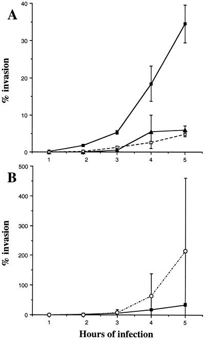
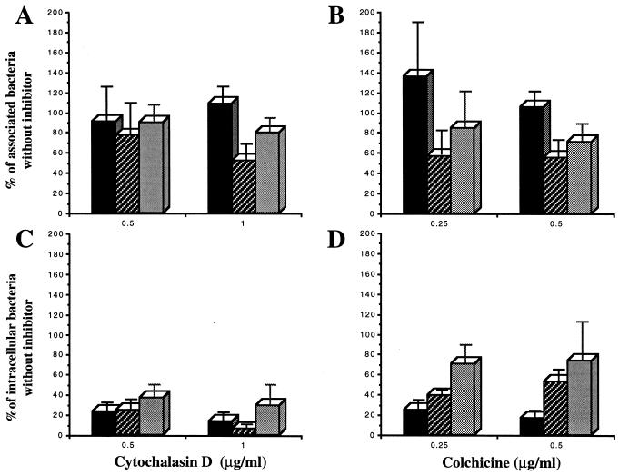
 , EIEC strain
E12860/0; ░⃞, EPEC strain E2348/69. Each value is the mean of at least
three separate experiments.
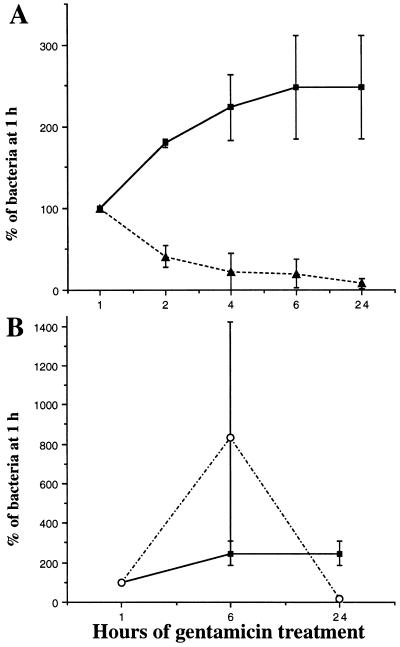
, EIEC strain
E12860/0; ░⃞, EPEC strain E2348/69. Each value is the mean of at least
three separate experiments.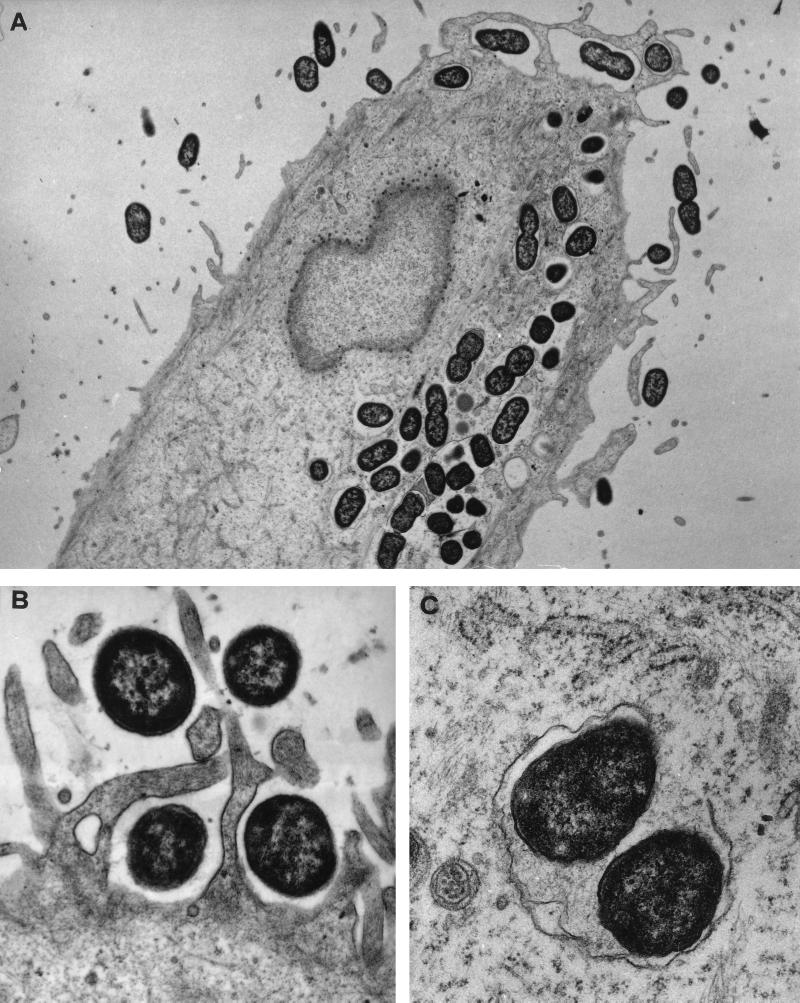
References
-
- Allaoui A, Sansonetti P J, Menard R, Barzu S, Mounier J, Phalipon A, Parsot C. MxiG, a membrane protein required for secretion of Shigellaspp. Ipa invasins: involvement in entry into epithelial cells and in intercellular dissemination. Mol Microbiol. 1995;17:461–470. - PubMed
-
- Andrade J R, Da Veiga V F, De Santa Rosa M R, Suassuna I. An endocytic process in HEp-2 cells induced by enteropathogenic Escherichia coli. J Med Microbiol. 1989;28:49–57. - PubMed
-
- Baudry B, Maurelli A T, Clerc P, Sadoff J C, Sansonetti P J. Localization of plasmid DNA loci necessary for the entry of Shigella flexneriinto Hela cells, and characterization of one locus encoding four immunogenic polypeptides. J Gen Microbiol. 1987;133:3409–3413. - PubMed
Publication types
MeSH terms
Substances
LinkOut - more resources
Full Text Sources
Other Literature Sources
Medical
