Comparative Study
. 1999 Sep;67(9):4563-9.
doi: 10.1128/IAI.67.9.4563-4569.1999.

Binding of rat and human surfactant proteins A and D to Aspergillus fumigatus conidia

Affiliations
Comparative Study

Binding of rat and human surfactant proteins A and D to Aspergillus fumigatus conidia

M J Allen et al. Infect Immun. 1999 Sep.

Abstract

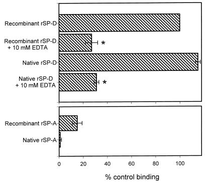
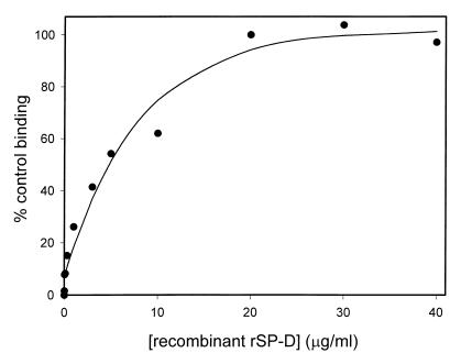
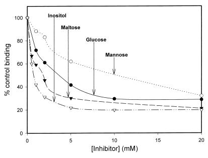
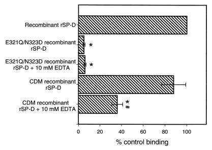
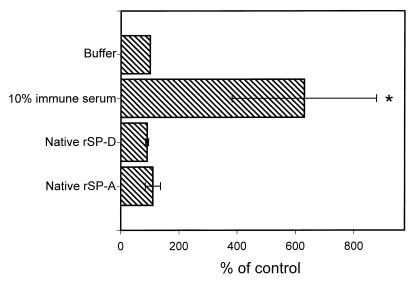
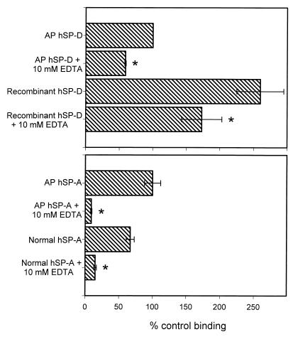
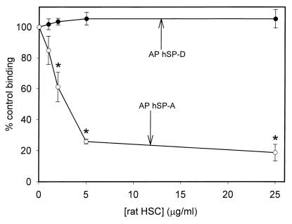
Surfactant proteins A (SP-A) and D (SP-D) are thought to play important roles in pulmonary host defense. We investigated the interactions of rat and human SP-A and SP-D with Aspergillus fumigatus conidia. Rat SP-D but not rat SP-A bound the conidia, and the binding was inhibited by EDTA, mannose, glucose, maltose, and inositol. Binding studies using a mutant recombinant rat SP-D with altered carbohydrate recognition but normal structural organization clearly established a role for the carbohydrate recognition domain in binding to conidia. However, neither rat SP-A nor SP-D increased the association of fluorescein isothiocyanate-labeled conidia with rat alveolar macrophages as determined by flow cytometry. Both human SP-A (isolated from normal and alveolar proteinosis lungs) and SP-D (recombinant protein and protein isolated from alveolar proteinosis lungs) bound the conidia. These data indicate that important differences exist between rat and human SP-A in binding to certain fungi. Human SP-A and SP-D binding to conidia was also examined in the presence of hydrophobic surfactant components (HSC), containing both the phospholipid and hydrophobic proteins of surfactant. We found that HSC inhibited but did not eliminate human SP-A binding to Aspergillus conidia. In contrast, the SP-D binding to conidia was unaffected by HSC. These findings indicate that SP-D plays a major role in the recognition of Aspergillus conidia in alveolar fluid.


Figures

FIG. 1
FIG. 1
Binding of native and recombinant rSP-A and rSP-D to A. fumigatus conidia. Aliquots of 2 × 10⁶ conidia were incubated with 20 μg of SP-A or SP-D/ml for 1 h at 25°C followed by washing and similar incubations with primary and secondary antibodies. For inhibition studies, the proteins were preincubated with EDTA for 15 min at 25°C and the EDTA-surfactant protein mixture was then added to the conidia. Binding was detected by flow cytometry and normalized to the mean fluorescent intensity of recombinant rSP-D (taken as 100% binding). Data are the averages ± standard errors of four independent experiments. ∗, P < 0.05 compared to binding without EDTA.
FIG. 2
FIG. 2
Dose response curve for recombinant rSP-D binding to A. fumigatus conidia. Aliquots of 2 × 10⁶ conidia were incubated with various concentrations of SP-D for 1 h at 25°C followed by washing and similar incubations with primary and secondary antibodies. Binding was detected by flow cytometry and normalized to the mean fluorescent intensity at 20 μg/ml. Data are the averages of duplicate analysis.
FIG. 3
FIG. 3
Inhibition of recombinant rSP-D binding to A. fumigatus conidia. Recombinant rSP-D (20 μg/ml) was incubated with various concentrations of mannose, glucose, maltose, and inositol for 15 min at 25°C. The SP-D-inhibitor mixture was then added to 2 × 10⁶ conidia and binding was allowed to progress for 1 h at 25°C, followed by washing and similar incubations with primary and secondary antibodies. Binding was detected by flow cytometry and normalized to the mean fluorescent intensity in the absence of inhibitor. Four independent experiments were performed for each inhibitor. The graph shows representative data for each inhibitor.
FIG. 4
FIG. 4
Binding of mutant recombinant rSP-D to A. fumigatus conidia. Aliquots of 2 × 10⁶ conidia were incubated with 20 μg of SP-D/ml for 1 h at 25°C followed by washing and similar incubations with primary and secondary antibodies. For inhibition studies, the proteins were preincubated with EDTA for 15 min at 25°C and the EDTA-surfactant protein mixture was then added to the conidia. Binding was detected by flow cytometry and normalized to the mean fluorescent intensity of wild-type recombinant rSP-D. ∗, P < 0.05 compared to the binding of wild-type recombinant rSP-D; #, P < 0.05 compared to mutant protein binding without EDTA. Data are the averages ± standard errors of four independent experiments. EDTA inhibition of wild-type recombinant rSP-D is shown in Fig. 1.
FIG. 5
FIG. 5
Association of A. fumigatus conidia with cultured rat alveolar macrophage cells. Samples of 9 × 10⁵ FITC-labeled conidia were incubated with native rSP-A (10 μg/ml), native rSP-D (10 μg/ml), 10% rabbit immune serum, or buffer for 30 min at 37°C. Following incubation, 3 × 10⁵ alveolar macrophage cells were added to the conidia and the mixture was incubated at 37°C for 1 h. The cells were then washed in buffer containing 10 mM EDTA and fixed with 2% paraformaldehyde in PBS for 30 min at room temperature. The cell association was determined by measuring the FITC fluorescence associated with the macrophages. The data were normalized to the buffer control and are the averages ± standard errors of four or five independent experiments. ∗, P < 0.05 compared to buffer control.
FIG. 6
FIG. 6
Binding of hSP-A and hSP-D to A. fumigatus conidia. Conidia (2 × 10⁶) were incubated with 20 μg of hSP-A or hSP-D/ml for 1 h at 25°C followed by washing and similar incubations with primary and secondary antibodies. For inhibition studies, the proteins were preincubated with EDTA for 15 min at 25°C and the EDTA-surfactant protein mixture was then added to the conidia. Binding was detected by flow cytometry and normalized to the mean fluorescent intensity of AP hSP-D. Data are the averages ± standard errors of three or four independent experiments. ∗, P < 0.05 compared to binding without EDTA.
FIG. 7
FIG. 7
Inhibition of AP hSP-A and AP hSP-D binding to A. fumigatus conidia by rat HSC. Purified AP hSP-A or AP-hSP-D (20 μg/ml) was incubated with various concentrations of rat HSC (based on total phospholipid content) for 15 min at 25°C. The surfactant protein-inhibitor mixture was then added to 2 × 10⁶ conidia and binding was allowed to progress for 1 h at 25°C, followed by washing and similar incubations with primary and secondary antibodies. Binding was detected by flow cytometry and normalized to the mean fluorescent intensity in the absence of inhibitor. The data are the averages ± standard errors of three independent experiments. ∗, P < 0.05 compared to binding in the absence of HSC.

References

    1. Allen M, Mason R, Harbeck R, Greene K, Smith B, Voelker D. Abstracts of the 1998 International Conference of the American Thoracic Society. Am. J. Respir. Crit. Care Med. 157:A865. 1998. Binding of rat SP-A and SP-D to Aspergillus spores.
    1. Benne C A, Benaissa-Trouw B, van Strijp J A, Kraaijeveld C A, van Iwaarden J F. Surfactant protein A, but not surfactant protein D, is an opsonin for influenza A virus phagocytosis by rat alveolar macrophages. Eur J Immunol. 1997;27:886–890. - PubMed
    1. Benne C A, Kraaijeveld C A, van Strijp J A, Brouwer E, Harmsen M, Verhoef J, van Golde L M, van Iwaarden J F. Interactions of surfactant protein A with influenza A viruses: binding and neutralization. J Infect Dis. 1995;171:335–341. - PubMed
    1. Crouch E C. Collectins and pulmonary host defense. Am J Respir Cell Mol Biol. 1998;19:177–201. - PubMed
    1. Hartshorn K, Chang D, Rust K, White M, Heuser J, Crouch E. Interactions of recombinant human pulmonary surfactant protein D and SP-D multimers with influenza A. Am J Physiol. 1996;271:L753–L762. - PubMed

Publication types

MeSH terms

Substances

LinkOut - more resources