. 1999 Sep;67(9):4586-93.
doi: 10.1128/IAI.67.9.4586-4593.1999.

Assessment of immunity to mycobacterial infection with luciferase reporter constructs

Affiliations

Assessment of immunity to mycobacterial infection with luciferase reporter constructs

V A Snewin et al. Infect Immun. 1999 Sep.

Abstract

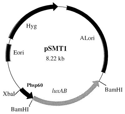
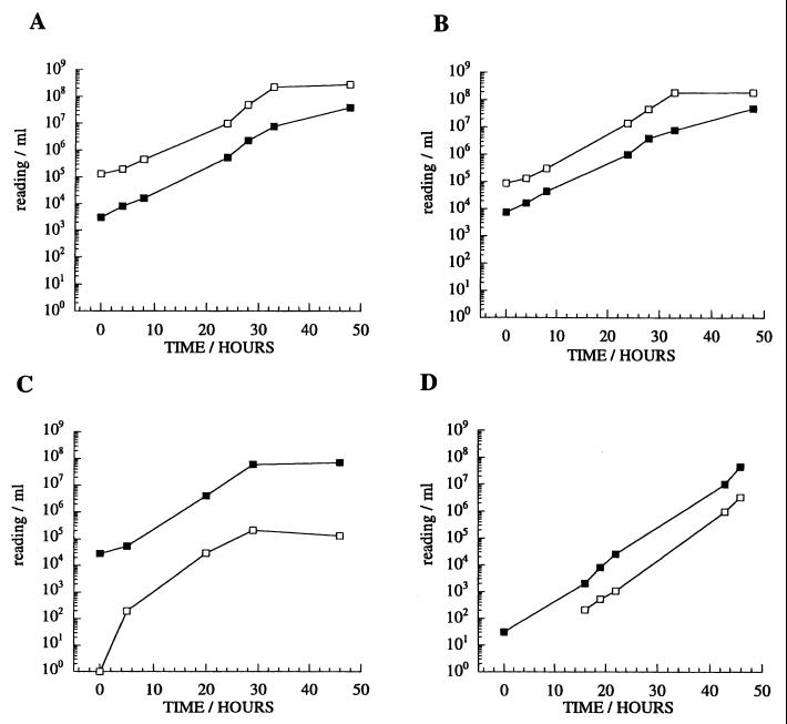
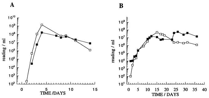
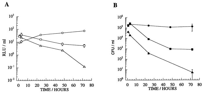
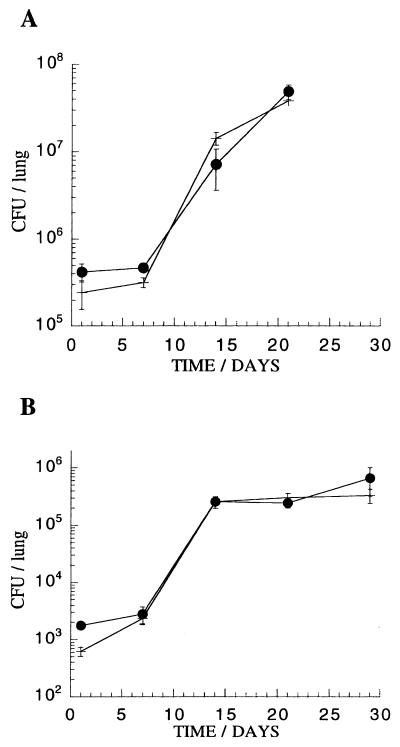
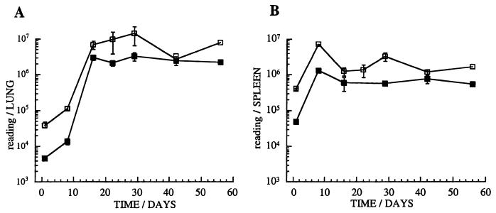
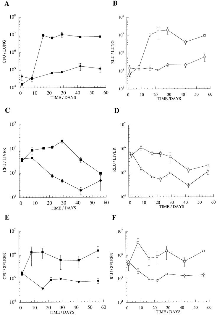
Protective immunity to mycobacterial infection is incompletely understood but probably involves the coordinated interaction of multiple cell types and cytokines. With the aim of developing assays that might provide a surrogate measure of protective immunity, we have investigated the use of recombinant mycobacteria carrying luciferase reporter enzymes to assess the effectiveness of antimycobacterial immunity in model systems. Measurement of luminescence was shown to provide a rapid and simple alternative to the counting of CFU as a means of monitoring mycobacterial viability. We describe optimization of a luciferase reporter strain of Mycobacterium tuberculosis and demonstrate its application for the study of mycobacterial interactions with host cells in tissue culture and the rapid assessment of vaccine efficacy in a murine model.


Figures

FIG. 1
FIG. 1
The construct pSMT1. Light production may be correlated with CFU. The reporter plasmid pSMT1 is a shuttle vector containing both an origin of replication for mycobacteria (ALori) and an E. coli origin of replication (Eori). Hygromycin (Hyg) is employed for antibiotic selection. The V. harveyi luxAB genes are under the control of the BCG hsp60 promoter (Phsp60). XbaI and BamHI cleavage sites are indicated.
FIG. 2
FIG. 2
Luminescence activities of M. smegmatis strains containing luciferase constructs. Liquid cultures were monitored over a time course of 48 h. (A) pSMT1 (V. harveyi luxA and luxB), (B) pSMT5 (X. luminescens luxA and luxB), (C) pSMT6 (firefly luc), (D) pLINT560 (V. harveyi luxA and luxB integrated in the chromosome; in this case, the RLU value at time point 0 was below the detection limits [approximately 100 RLUs]). Open symbols, RLUs per milliliter; closed symbols, CFU per milliliter.
FIG. 3
FIG. 3
Comparison of luciferase expression in M. smegmatis (A) and M. tuberculosis H37Rv (B) strains containing the plasmid pSMT1. RLU and CFU readings were taken during log-phase and stationary-phase growth. Background readings obtained from wild-type mycobacteria and mycobacteria transfected with a control plasmid were routinely approximately 100 RLUs. Open symbols, RLUs per milliliter; closed symbols, CFU per milliliter.
FIG. 4
FIG. 4
Mycobacterial infection of J774A.1 cells. Viability was determined by RLU (A) and CFU (B) readings. MOI was approximately 1. Circles, M. tuberculosis H37Rv/pSMT1; squares, BCG/pSMT1; triangles, M. smegmatis/pSMT1. Such infections generally yield between 1 and 10% infected cells, with approximately 3 to 5 bacteria per infected cell. The means and standard deviations (error bars) are shown for three wells at each time point.
FIG. 5
FIG. 5
Comparison of growth rate in C57BL/6 mice of M. tuberculosis H37Rv/pSMT1 (closed circles) with M. tuberculosis H37Rv/pSMT3, a control plasmid without the luciferase luxAB genes (cross). (A) High inoculum (3.4 × 10⁵ and 3.6 × 10⁵ bacteria, respectively, per mouse) and (B) low inoculum (3.4 × 10³ and 3.6 × 10³ bacteria, respectively, per mouse) following i.v. challenge. The means and standard deviations (error bars) are shown for groups of three mice at each time point.
FIG. 6
FIG. 6
A murine model of infection with luminescent M. tuberculosis. C57BL/6 mice were infected with 5 × 10⁵ M. tuberculosis H37Rv/pSMT1 bacteria. The relationship between RLUs (open squares) and CFU (closed squares) is illustrated for homogenates of lung (A) and spleen (B) in this experiment (three mice per group). RLU readings of uninfected splenocytes prepared in the same way showed background luminescence (100 RLUs). The means and standard deviations (error bars) are shown for groups of three mice at each time point.
FIG. 7
FIG. 7
A vaccination model of murine infection with luminescent M. tuberculosis. The course of M. tuberculosis infection was monitored by measurements of CFU and RLUs in homogenates of lung (A and B), liver (C and D), and spleen (E and F) of mice immunized by BCG vaccination (circles) and of naive control mice (squares). The means and standard deviations (error bars) are shown for groups of three mice at each time point.

References

    1. Andrew P W, Roberts I S. Construction of a bioluminescent mycobacterium and its use for assay of antimycobacterial agents. J Clin Microbiol. 1993;31:2251–2254. - PMC - PubMed
    1. Arain T M, Resconi A E, Hickey M J, Stover K. Bioluminescence screening in vitro (Bio-siv) assays for high-volume antimycobacterial drug discovery. Antimicrob Agents Chemother. 1996;40:1536–1541. - PMC - PubMed
    1. Arain T M, Resconi A E, Singh D C, Stover K. Reporter gene technology to assess activity of antimycobacterial agents in macrophages. Antimicrob Agents Chemother. 1996;40:1542–1544. - PMC - PubMed
    1. Barry M A, Lai W C, Johnston S A. Protection against mycoplasma infection using expression-library immunization. Nature. 1995;377:632–635. - PubMed
    1. Carriere C, Riska P, Zimhony O, Kriakov J, Bardarov S, Burns J, Chan J, Jacobs W R., Jr Conditionally replicating luciferase reporter phages: improved sensitivity for rapid detection and assessment of drug susceptibility of Mycobacterium tuberculosis. J Clin Microbiol. 1997;35:3232–3239. - PMC - PubMed

Publication types

MeSH terms