. 1999 Sep;67(9):4613-9.
doi: 10.1128/IAI.67.9.4613-4619.1999.

Pseudomonas aeruginosa exoenzyme S stimulates murine lymphocyte proliferation in vitro

Affiliations

Pseudomonas aeruginosa exoenzyme S stimulates murine lymphocyte proliferation in vitro

N G Barclay et al. Infect Immun. 1999 Sep.

Abstract

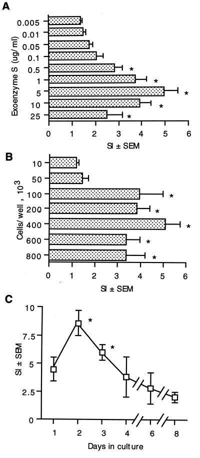
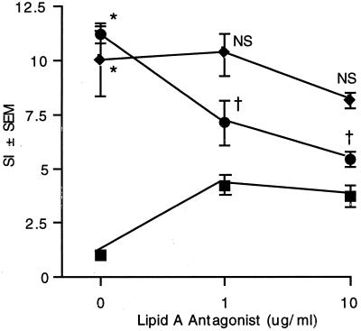
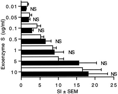
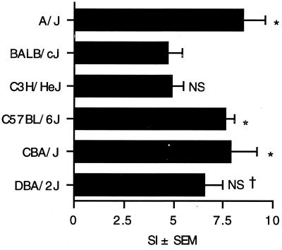
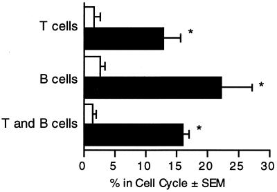
The exuberant immunoinflammatory response that is associated with Pseudomonas aeruginosa infection is the major source of the morbidity and mortality in cystic fibrosis (CF) patients. Previous studies have established that an exoproduct of P. aeruginosa (exoenzyme S) is a mitogen for human T lymphocytes and activates a larger percentage of T cells than most superantigens, which may contribute to the immunoinflammatory response. An animal model would facilitate studies of the pathophysiologic consequences of this activation. As a first step toward developing an animal model, the murine lymphocyte response to exoenzyme S was examined. When stimulated with exoenzyme S, splenocytes isolated from naive mice entered S phase and proliferated. The optimum response occurred after 2 to 3 days in culture, at 4 x 10(5) cells per well and 5.0 micrograms of exoenzyme S per ml. The response was not due to lipopolysaccharide, since Rhodobacter sphaeroides lipid A antagonist did not block the response. Other preparations of exoenzyme S stimulated lymphocyte proliferation, since the response to recombinant exoenzyme S (rHisExo S) cloned from strain 388 was similar to the response to exoenzyme S from strain DG1. There was evidence that genetic variability influenced the response, since A/J, CBA/J, and C57BL/6 mice were high responders and BALB/cJ mice were low responders following stimulation with exoenzyme S. Both splenic T and B lymphocytes entered the cell cycle in response to exoenzyme S. Thus, murine lymphocytes, like human lymphocytes, respond to P. aeruginosa exoenzyme S, which supports the development of a murine model that may facilitate our understanding of the role that exoenzyme S plays in the pathogenesis of P. aeruginosa infections in CF patients.


Figures

FIG. 1
FIG. 1
Conditions required for murine splenocyte proliferation in response to stimulation with exoenzyme S. (A) Splenocytes from C3H/HeJ mice (4 × 10⁵/well) were cultured with 0.005 to 25 μg of exoenzyme S per ml for 3 days. *, P < 0.05 compared to splenocytes stimulated with 0.005 μg of exoenzyme S per ml. [3H]TdR incorporation in unstimulated splenocytes = 2.7 (± 0.4) × 103 cpm; in response to 1 μg of Staphylococcus enterotoxin B per ml, SI = 9.1 ± 1.0. (B) Splenocytes (1 × 10⁴ to 8 × 10⁵/well) from C3H/HeJ mice were cultured with 1 μg of exoenzyme S per ml for 3 days. *, P < 0.05 compared to 10 × 103 splenocytes per well. (C) Splenocytes from C3H/HeJ mice (4 × 10⁵/well) were cultured with 1 μg of exoenzyme S per ml for 1 to 8 days. *, P < 0.05 compared to splenocytes stimulated for 8 days. Values represent the mean SI ± SEM from eight different experiments.
FIG. 2
FIG. 2
The proliferative response of splenocytes to exoenzyme S is not due to contaminating LPS. Splenocytes (4 × 10⁵/well) from LPS-responsive DBA/2J mice were stimulated with exoenzyme S (⧫) (1 μg/ml) or P. aeruginosa LPS (●) (1 μg/ml) or left unstimulated (■) in the presence of various concentrations of R. sphaeroides lipid A antagonist. Values represent the mean SI of [3H]TdR incorporation ± SEM from five experiments. *, P < 0.05 compared to unstimulated splenocytes. NS, not significant compared to exoenzyme S without lipid A antagonist. †, P < 0.05 compared to P. aeruginosa LPS without lipid A antagonist. [3H]TdR incorporation in unstimulated splenocytes = 3.1 (± 0.5) × 103 cpm.
FIG. 3
FIG. 3
Different preparations of exoenzyme S stimulate splenocytes to proliferate. Splenocytes (4 × 10⁵/well) from DBA/2J mice were cultured for 3 days in the presence of exoenzyme S purified from P. aeruginosa DG1 (□) or rHisExo S expressed in E. coli BL21(DE3) (■). Values represent the mean SI of [3H]TdR incorporation ± SEM from five experiments. NS, not significant compared to the corresponding concentration of exoenzyme S purified from P. aeruginosa DG1.
FIG. 4
FIG. 4
Splenocyte proliferation in different strains of inbred mice in response to exoenzyme S. Splenocytes (4 × 10⁵/well) from A/J, BALB/cJ, C3H/HeJ, C57BL/6J, CBA/J, or DBA/2J mice were cultured for 3 days in medium alone or with exoenzyme S (5 μg/ml). The bars represent the mean SI of [3H]TdR incorporation ± SEM from four mice. *, P < 0.05 compared to BALB/cJ and C3H/HeJ mice. NS, not significant compared to BALB/cJ mice. †, not significant compared to A/J, C57BL/6J, or CBA/J mice.
FIG. 5
FIG. 5
B cells and T cells enter the cell cycle in response to stimulation by P. aeruginosa exoenzyme S. Splenocytes (4 × 10⁵/well) were cultured with medium alone (open bars) or 5 μg of exoenzyme S per ml (solid bars) for 3 days. Cells were labeled with either anti-Thy 1.(T cells), anti-B220 (B cells), or no antibody (T and B cells) and analyzed for DNA content by propidium iodide labeling with flow cytometry. The percentage of cells in the cell cycle was determined by CellQuest analysis. The bars represent the mean percentage of cells in the cycle ± SEM from five different experiments. *, P < 0.05 compared to the corresponding unstimulated group.

References

    1. Bach M, Hoffenbach A. Strain-dependent protective effect of adult thymectomy on murine infection by Mycobacterium lepraemurium. Clin Exp Immunol. 1987;68:521–527. - PMC - PubMed
    1. Boat T F, Welsh M J, Beaudet A L. Cystic fibrosis. In: Scriver C R, Beadet A L, Sly W S, Valle D, editors. The metabolic basis of inherited disease. New York, N.Y: McGraw-Hill; 1989. pp. 2649–2680.
    1. Boom W H, Liebster L, Abbas A K, Titus R G. Patterns of cytokine secretion in murine leishmaniasis: correlation with disease progression or resolution. Infect Immun. 1990;58:3863–3870. - PMC - PubMed
    1. Bruno T F, Syme R M, Buser D E, Woods D E, Mody C H. Pseudomonas aeruginosa exoenzyme S is a mitogen, but not a superantigen for adult T lymphocytes. Infect Immun. 1998;66:3072–3079. - PMC - PubMed
    1. Bruno T F, Woods D E, Mody C H. Recombinant Pseudomonas exoenzyme S and exoenzyme S from Pseudomonas aeruginosa strain DG1 stimulate T lymphocytes to proliferate. Can J Microbiol. 1999;45:1–5. .. - PubMed

Publication types

MeSH terms