. 1999 Sep;67(9):4700-7.
doi: 10.1128/IAI.67.9.4700-4707.1999.

Preliminary characterization of a Mycobacterium abscessus mutant in human and murine models of infection

Affiliations

Preliminary characterization of a Mycobacterium abscessus mutant in human and murine models of infection

T F Byrd et al. Infect Immun. 1999 Sep.

Abstract

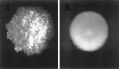
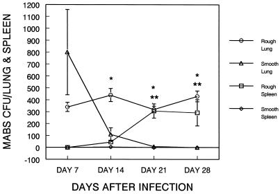
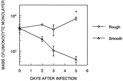
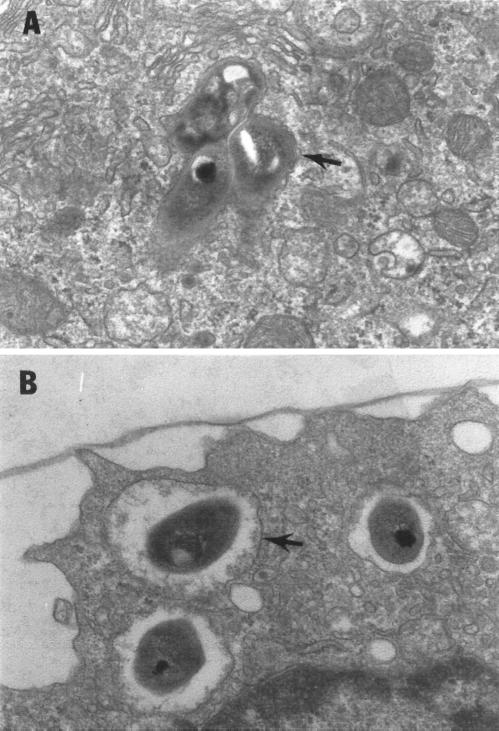
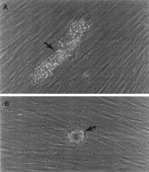
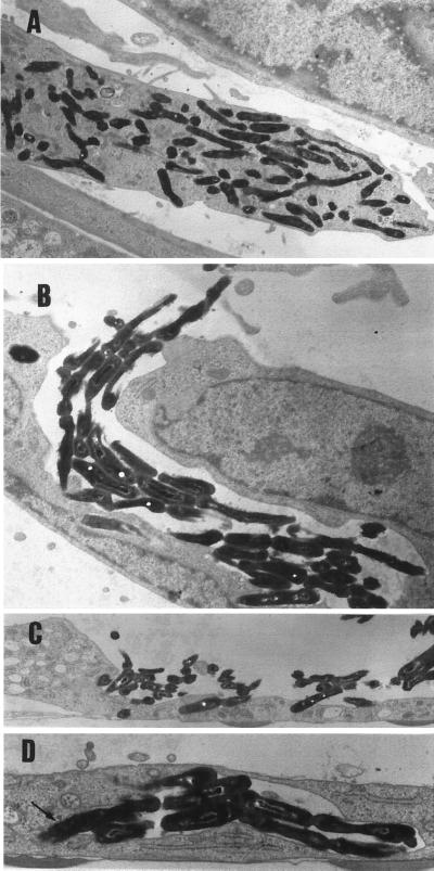
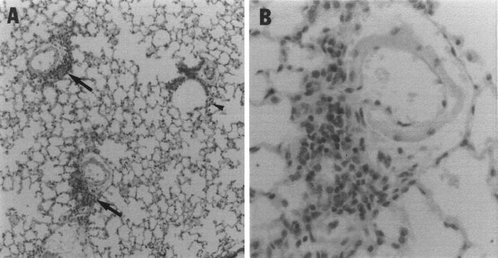
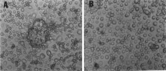
The ability to persist in the host after the establishment of infection is an important virulence determinant for mycobacteria. Mycobacterium abscessus is a rapidly growing mycobacterial species which causes a variety of clinical syndromes in humans. We have obtained a rough, wild-type human clinical isolate of M. abscessus (M. abscessus-R) and a smooth, attenuated mutant (M. abscessus-S) which spontaneously dissociated from the clinical isolate. We have found that M. abscessus-R is able to persist and multiply in a murine pulmonary infection model in contrast to M. abscessus-S, which is rapidly cleared. To understand the basis for this difference, we characterized the behavior of these variants in human tissue culture models of infection. M. abscessus-R is able to persist and multiply in human monocytes, while M. abscessus-S is deficient in this ability. Both of these variants are phagocytized by human monocytes. M. abscessus-R resides in a phagosome typical for pathogenic mycobacteria with a tightly adherent phagosomal membrane. In contrast, M. abscessus-S resides in a "loose" phagosome with the phagosomal membrane separated from the bacterial cell wall. Both M. abscessus variants also have distinctive growth patterns in a recently described fibroblast-mycobacterium microcolony assay, with M. abscessus-R exhibiting growth characteristics similar to those previously reported for virulent M. tuberculosis and M. abscessus-S exhibiting growth characteristics similar to those previously reported for avirulent M. tuberculosis. In both the monocyte infection assay and the murine pulmonary infection model, numerous infected mononuclear phagocyte aggregates develop at sites of M. abscessus-R infection, but are absent with M. abscessus-S infection. We conclude that a mutation has occurred in the M. abscessus-S variant which has altered the ability of this organism to persist and multiply in host cells and that this may be related to the phenotypic changes we have observed in our tissue culture models of infection.


Figures

FIG. 1
FIG. 1
Morphologic appearance of M. abscessus-R (left) and M. abscessus-S (right) colonies. These are 7-day-old colonies on 7H11 agar.
FIG. 2
FIG. 2
In a murine pulmonary model of infection, M. abscessus-R persists in both the lung and spleen and disseminates to the spleen, whereas M. abscessus-S is rapidly cleared and does not disseminate. SCID mice (n = 4) were intratracheally inoculated with 104 M. abscessus-R or M. abscessus-S organisms. At the indicated time intervals, the mice were sacrificed and the total lung and spleen CFU values were determined. The data represent the mean ± the standard deviation. ∗, M. abscessus-R versus M. abscessus-S in the lung (P < 0.05); ∗∗, M. abscessus-R versus M. abscessus-S in the spleen (P < 0.05) (t test).
FIG. 3
FIG. 3
M. abscessus-R persists and multiplies in human monocyte monolayers in contrast to M. abscessus-S. Human monocyte monolayers were infected with 2.5 × 10⁵ M. abscessus-R or M. abscessus-S for 6 h. After being washed, cell lysates and supernatants were plated for CFU at the indicated intervals. CFU in supernatants were less than 5% of the CFU in lysates at all points. Data are the mean of duplicate determinations of two separate experiments. ∗, M. abscessus-R versus M. abscessus-S (P < 0.002; t test).
FIG. 4
FIG. 4
M. abscessus variants are phagocytized by human monocytes and reside in phagosomes with differing morphology. Human monocyte monolayers were incubated with 106 M. abscessus-R or M. abscessus-S for 1 h, followed by washing and an additional 30 min of incubation to allow for internalization of bacteria. Electron microscopy was then performed. Fifteen consecutive phagosomes were identified for each group, containing one to four bacteria per monocyte. All bacteria were within monocytes. (A) Three M. abscessus-R in a phagosome typical for pathogenic mycobacteria with a tightly adherent phagosomal membrane (arrow). (B) Three M. abscessus-S organisms in separate “loose” phagosomes with the phagosomal membrane separated from the bacterial cell wall (arrow). Magnification, ×12,000.
FIG. 5
FIG. 5
M. abscessus variants form distinctive microcolonies in the fibroblast-mycobacterium microcolony assay. The microscopic appearance of 2-day-old, unfixed, unstained M. abscessus growing in human fibroblast monolayers which had been overlaid with agar-tissue culture medium can be seen. (A) M. abscessus-R. (B) M. abscessus-S. Magnification, ×200.
FIG. 6
FIG. 6
M. abscessus-R multiplies extensively within human fibroblasts and secondarily invades cells after primary infection. (A) Numerous M. abscessus-R in fibroblast 2 days after infection. Magnification, ×4,900. (B) M. abscessus-R cord effacing and secondarily invading fibroblast. Magnification, ×7,700. (C) M. abscessus-R effacing and penetrating the surface of a fibroblast. Magnification, ×4,900. (D) M. abscessus-R cord that has been internalized by a fibroblast with bacteria becoming tightly associated with phagosomal membrane (arrow). Magnification, ×9,800. No secondary invasion of fibroblasts by M. abscessus-S was identified.
FIG. 7
FIG. 7
The lungs of SCID mice infected with M. abscessus-R contain numerous inflammatory foci which are absent in the lungs of M. abscessus-S-infected mice. (A) Seven days after infection with M. abscessus-R. Arrows indicate prominent perivascular inflammatory infiltrates. The arrowhead points to a small number of peribronchiolar inflammatory cells. The tissue was stained with hematoxylin and eosin and photographed at ×25. (B) Enlargement of perivascular inflammatory infiltrate seen in panel A. Magnification, ×100.
FIG. 8
FIG. 8
M. abscessus-R infection of human monocytes is associated with the development of monocyte aggregates. Human monocyte monolayers were infected with the M. abscessus variants as in Fig. 3. After 3 days, supernatants were removed and the monolayers were examined by trypan blue exclusion. (A) Monolayers infected with M. abscessus-R form dense cellular aggregates. (B) Cellular aggregates are completely lacking with M. abscessus-S. Magnification (both panels), ×100.

References

    1. Armstrong J A, Hart P D A. Phagosome-lysosome interactions in cultured macrophages infected with virulent tubercle bacilli. J Exp Med. 1975;142:1–16. - PMC - PubMed
    1. Barker L P, George K M, Falkow S, Small P L. Differential trafficking of live and dead Mycobacterium marinum organisms in macrophages. Infect Immun. 1997;65:1497–1504. - PMC - PubMed
    1. Beaman, B. Personal communication.
    1. Behling C A, Bennett B, Takayama K, Hunter R L. Development of a trehalose 6,6′-dimycolate model which explains cord formation by Mycobacterium tuberculosis. Infect Immun. 1993;61:2296–2303. - PMC - PubMed
    1. Belisle J T, McNeil M R, Chatterjee D, Inamine J M, Brennan P J. Expression of the core lipopeptide of the glycopeptidolipid surface antigens in rough mutants of Mycobacterium avium. J Biol Chem. 1993;268:10510–10516. - PubMed

Publication types

MeSH terms