. 1999 Sep;67(9):4757-63.
doi: 10.1128/IAI.67.9.4757-4763.1999.

T-Cell responses to Treponema pallidum subsp. pallidum antigens during the course of experimental syphilis infection

Affiliations

T-Cell responses to Treponema pallidum subsp. pallidum antigens during the course of experimental syphilis infection

T W Arroll et al. Infect Immun. 1999 Sep.

Abstract

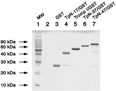
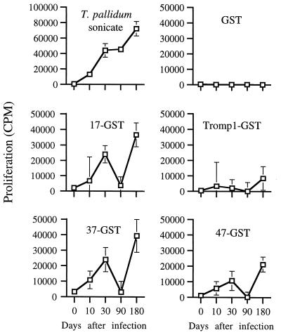
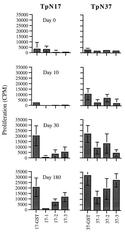
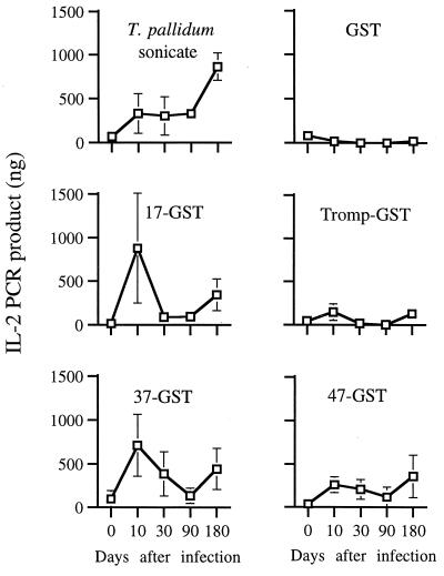
In this study we describe the development of the T-cell response to a panel of Treponema pallidum antigens over the course of syphilis infection in the rabbit and determine whether these antigens induce the expression of Th1 cytokines. It was determined that the membrane proteins TpN17 and TpN47, as well as the endoflagellar sheath protein TpN37, induce strong proliferation responses through most of syphilis infection; Tromp1 induced only weak proliferative responses. An unexpected drop in proliferative response to these antigens at day 90 of infection, followed by a dramatic increase in response at day 180, suggests that there may be a secondary dissemination of T. pallidum which induces a recall response. Crude epitope mapping of TpN17 and TpN37 showed that multiple epitopes may be present on both antigens, which is likely a contributing factor in the immunodominance of these antigens. The T-cell response to the TpN37 molecule shows acquisition of newly recognized epitopes during the course of infection. Sonicated T. pallidum was found to induce the expression of interleukin-2 (IL-2) and gamma interferon and not IL-10 mRNA, showing that the general T-cell response to T. pallidum antigens in syphilis infection is biased towards the Th1 phenotype. Of the antigens tested, TpN37 appears to contribute the most to the Th1 cytokine response and therefore may play a key role in the clearance of T. pallidum from lesions.


Figures

FIG. 1
FIG. 1
GST fusion proteins were purified by either glutathione-agarose or nickel affinity chromatography as described in the Materials and Methods. Ten micrograms of each protein were resolved on an SDS–12.5% PAGE gel and stained with Coomassie blue.
FIG. 2
FIG. 2
Splenocyte proliferative responses to T. pallidum-GST fusion proteins. Splenic lymphocytes from normal rabbits (day 0) (n = 9) and from rabbits infected with T. pallidum for 10 (n = 11), 30 (n = 8), 90 (n = 8), or 180 (n = 4) days were tested for proliferative responses to the panel of recombinant T. pallidum antigens. The data points represent the geometric means of the proliferative measurements of each group of rabbits ± the standard error of the mean (SEM). The background proliferative response (no antigen) for each condition was less than 2,500 cpm. Data from at least two groups of infected rabbits are shown for each time point except for day 180, which is from one group of infected rabbits.
FIG. 3
FIG. 3
Mapping of epitopes recognized by splenic lymphocytes. Truncated fragments of TpN17 and TpN37 expressed as GST fusion proteins were tested for proliferative responses in splenic lymphocytes from normal rabbits (day 0) and a group of rabbits infected with T. pallidum for 10, 30, and 180 days (n = 4 for each time point). The data points represent the geometric means of the proliferation measurements of each group of rabbits ± the SEM.
FIG. 4
FIG. 4
IFN-γ mRNA expression in response to T. pallidum-GST fusion proteins. Total RNA was isolated from splenocytes cultured with antigen; IFN-γ message was measured by RT-PCR. Data points represent the means ± the SEM of values measured in groups of four animals infected at the same time for all time points except day 30 (three rabbits).
FIG. 5
FIG. 5
IL-2 mRNA expression in response to T. pallidum-GST fusion proteins. Total RNA was isolated from splenocytes cultured with antigens; IL-2 message was measured by RT-PCR. Data points represent the means ± the SEM of values measured in groups of four animals infected at the same time for all time points except day 30 (three rabbits).

References

    1. Akins D R, Robinson E, Shevchenko D, Elkins C, Cox D L, Radolf J D. Tromp1, a putative rare outer membrane protein, is anchored by an uncleaved signal sequence to the Treponema pallidum cytoplasmic membrane. J Bacteriol. 1997;179:5076–5086. - PMC - PubMed
    1. Bailey M J, Cockayne A, Penn C W. Production of murine monoclonal antibodies to the major axial filament polypeptide of Treponema pallidum. J Gen Microbiol. 1987;133:1805–1813. - PubMed
    1. Baker-Zander S, Sell S. A histopathologic and immunologic study of the course of syphilis in the experimentally infected rabbit. Demonstration of long-lasting cellular immunity. Am J Pathol. 1980;101:387–413. - PMC - PubMed
    1. Baker-Zander S A, Fohn M J, Lukehart S A. Development of cellular immunity to individual soluble antigens of Treponema pallidum during experimental syphilis. J Immunol. 1988;141:4363–4369. - PubMed
    1. Bishop N H, Miller J N. Humoral immunity in experimental syphilis. I. The demonstration of resistance conferred by passive immunization. J Immunol. 1976;117:191–196. - PubMed

MeSH terms