. 1999 Sep;67(9):4902-7.
doi: 10.1128/IAI.67.9.4902-4907.1999.

Use of genetically manipulated strains of Clostridium perfringens reveals that both alpha-toxin and theta-toxin are required for vascular leukostasis to occur in experimental gas gangrene

Affiliations

Use of genetically manipulated strains of Clostridium perfringens reveals that both alpha-toxin and theta-toxin are required for vascular leukostasis to occur in experimental gas gangrene

D M Ellemor et al. Infect Immun. 1999 Sep.

Abstract

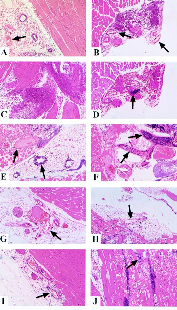
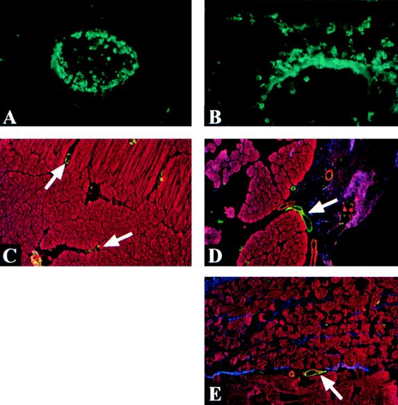
A hallmark of gas gangrene (clostridial myonecrosis) pathology is a paucity of leukocytes infiltrating the necrotic tissue. The cause of this paucity most likely relates to the observation of leukocyte aggregates at the border of the area of tissue necrosis, often within the microvasculature itself. Infecting mice with genetically manipulated strains of Clostridium perfringens type A (deficient in either alpha-toxin or theta-toxin production) resulted in significantly reduced leukocyte aggregation when alpha-toxin was absent and complete abrogation of leukocyte aggregation when theta-toxin was absent. Thus, both alpha-toxin and theta-toxin are necessary for the characteristic vascular leukostasis observed in clostridial myonecrosis.


Figures

FIG. 1
FIG. 1
Histological analysis of tissue injected with various strains of C. perfringens type A. Sections were stained with hematoxylin and eosin unless otherwise stated. Arrows indicate the positions of blood vessels or bacterium-leukocyte aggregates. (A) Inoculum containing PBS only. There is little leukocyte infiltration into the tissue, and there is no leukocyte accumulation within small venules. (B, C, and D) Heat-killed C. perfringens inoculum. There is little or no leukocyte accumulation within blood vessels at 4 h postinjection (panel B). Extensive leukocyte infiltration into the infected tissue is apparent by 8 h (panel C). Infiltrating leukocytes are observed to surround and penetrate bacterial aggregates by 4 h (analyzed by using gram-stained serial section) (panel D). (E and F) Wild-type C. perfringens inoculum. Profound leukocyte accumulation and thrombosis are observed in vessels collected at 4 h (panel E) and 8 h (panel F), with very little leukocyte infiltration into surrounding tissue. (G and H) Theta-toxin-deficient C. perfringens inoculum. Little or no leukocyte accumulation is present within blood vessels at 4 h although blood vessel thrombosis is prominent (panel G). Significant leukocyte infiltration of surrounding tissue is observed at 8 h (panel H), although leukocytes rarely colocalize with bacterial aggregates. (I and J) Alpha-toxin-deficient C. perfringens inoculum. Occasional leukocyte accumulation is observed within blood vessels at 8 h (panel I); blood vessel thrombosis is rarely observed. Significant leukocyte infiltration of surrounding tissue is apparent by 12 h (panel J); penetration and clearance of the aggregates occur but to a reduced degree compared to that observed in tissues injected with heat-killed C. perfringens cells.
FIG. 2
FIG. 2
Immunofluorescence staining of Mac-1 and ICAM-1. (A and B) Mac-1 staining. Accumulation of leukocytes within blood vessels is marked for tissue injected with wild-type C. perfringens inocula (panel A) compared to blood vessels in tissue injected with heat-killed C. perfringens inocula (panel B), which remain free of leukocyte build-up. Polarized staining of the blood vessel wall observed in the heat-killed C. perfringens-injected tissue (panel B) is most likely attributable to shedding of Mac-1 by leukocytes during direct transendothelial migration. (C, D, and E) ICAM-1 staining. The apical surface of endothelial cells stained strongly for ICAM-1 (green) in tissues injected with PBS alone (panel C) or with heat-killed (panel D) and wild-type (panel E) C. perfringens cells. C. perfringens cells (blue) and muscle fibers (red) were also visualized in these tissues by using antibody conjugates. The intensity of ICAM-1 staining in tissue injected with heat-killed or wild-type C. perfringens cells was observed to be consistently brighter than that in tissue injected with PBS alone, although the difference was not highly significant. No difference in ICAM-1 staining intensities between tissue samples injected with either live or heat-killed C. perfringens cells was observed.

References

    1. Awad M, Bryant A, Stevens D, Rood J. Virulence studies on chromosomal α-toxin and θ-toxin mutants constructed by allelic exchange provide genetic evidence for the essential role of α-toxin in Clostridium perfringens-mediated gas gangrene. Mol Microbiol. 1995;15:191–202. - PubMed
    1. Awad M, Rood J. Isolation of α-toxin, θ-toxin and κ-toxin mutants of Clostridium perfringens by Tn916 mutagenesis. Microb Pathog. 1997;22:275–284. - PubMed
    1. Bannam T, Rood J. Clostridium perfringens-Escherichia coli shuttle vectors that carry single antibiotic resistance determinants. Plasmid. 1992;229:233–235. - PubMed
    1. Bryant A, Bergstrom R, Zimmerman G, Salyer J, Hill H, Tweten R, Sato H, Stevens D. Clostridium perfringens invasiveness is enhanced by effects of theta toxin upon PMNL structure and function: the roles of leukocytotoxicity and expression of CD11/CD18 adherence glycoprotein. FEMS Immunol Med Microbiol. 1993;7:321–336. - PubMed
    1. Bryant A, Stevens D. Phospholipase C and perfringolysin O from Clostridium perfringens upregulate endothelial cell-leukocyte adherence molecule 1 and intercellular leukocyte adherence molecule 1 expression and induce interleukin-8 synthesis in cultured human umbilical vein endothelial cells. Infect Immun. 1996;64:358–362. - PMC - PubMed

Publication types

Substances

LinkOut - more resources