

Role of complement receptors in uptake of Mycobacterium avium by macrophages in vivo: evidence from studies using CD18-deficient mice
- PMID: 10456949
- PMCID: PMC96827
- DOI: 10.1128/IAI.67.9.4912-4916.1999
Role of complement receptors in uptake of Mycobacterium avium by macrophages in vivo: evidence from studies using CD18-deficient mice
Abstract
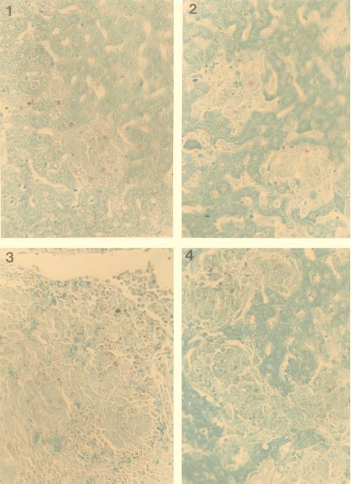
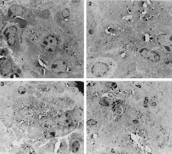
Mycobacterium avium is an intracellular pathogen that has been shown to invade macrophages by using complement receptors in vitro, but mycobacteria released from one cell can enter a second macrophage by using receptors different from complement receptors. Infection of CD18 (beta(2) integrin) knockout mice and the C57 BL/6 control mice led to comparable levels of tissue infection at 1 day, 2 days, 1 week, and 3 weeks following administration of bacteria. A histopathological study revealed similar granulomatous lesions in the two mouse strains, with comparable numbers of organisms. In addition, transmission electron microscopy of spleen tissues from both strains of mice showed bacteria inside macrophages. Our in vivo findings support the hypothesis that M. avium in the host is likely to use receptors other than CR3 and CR4 receptors to enter macrophages with increased efficiency.
Figures
References
-
- Bermudez L E, Stevens P, Kolonoski P, Wu M, Young L S. Treatment of disseminated Mycobacterium avium complex infection in mice with recombinant interleukin-2 and tumor necrosis factor. J Immunol. 1989;143:2996–3002. - PubMed
Publication types
MeSH terms
Substances
Grants and funding
LinkOut - more resources
Full Text Sources
