. 1999 Sep 1;519 Pt 2(Pt 2):347-60.
doi: 10.1111/j.1469-7793.1999.0347m.x.

ATP-sensitive K+ channel openers prevent Ca2+ overload in rat cardiac mitochondria

Affiliations

ATP-sensitive K+ channel openers prevent Ca2+ overload in rat cardiac mitochondria

E L Holmuhamedov et al. J Physiol. .

Abstract

1. Mitochondrial dysfunction, secondary to excessive accumulation of Ca2+, has been implicated in cardiac injury. We here examined the action of potassium channel openers on mitochondrial Ca2+ homeostasis, as these cardioprotective ion channel modulators have recently been shown to target a mitochondrial ATP-sensitive K+ channel. 2. In isolated cardiac mitochondria, diazoxide and pinacidil decreased the rate and magnitude of Ca2+ uptake into the mitochondrial matrix with an IC50 of 65 and 128 microM, respectively. At all stages of Ca2+ uptake, the potassium channel openers depolarized the mitochondrial membrane thereby reducing Ca2+ influx through the potential-dependent mitochondrial uniporter. 3. Diazoxide and pinacidil, in a concentration-dependent manner, also activated release of Ca2+ from mitochondria. This was prevented by cyclosporin A, an inhibitor of Ca2+ release through the mitochondrial permeability transition pore. 4. Replacement of extramitochondrial K+ with mannitol abolished the effects of diazoxide and pinacidil on mitochondrial Ca2+, while the K+ ionophore valinomycin mimicked the effects of the potassium channel openers. 5. ATP and ADP, which block K+ flux through mitochondrial ATP-sensitive K+ channels, inhibited the effects of potassium channel openers, without preventing the action of valinomycin. 6. In intact cardiomyocytes, diazoxide also induced mitochondrial depolarization and decreased mitochondrial Ca2+ content. These effects were inhibited by the mitochondrial ATP-sensitive K+ channel blocker 5-hydroxydecanoic acid. 7. Thus, potassium channel openers prevent mitochondrial Ca2+ overload by reducing the driving force for Ca2+ uptake and by activating cyclosporin-sensitive Ca2+ release. In this regard, modulators of an ATP-sensitive mitochondrial K+ conductance may contribute to the maintenance of mitochondrial Ca2+ homeostasis.


Figures
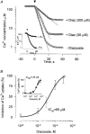
Figure 1
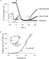
Figure 1. Potassium channel openers reduce Ca2+ uptake in isolated cardiac mitochondria
A, Ca2+ concentration within the mitochondrial suspension, illustrating the effect of 30 and 300 μm diazoxide (Diaz) on slowing the rate of mitochondrial Ca2+ uptake, and decreasing the amount of accumulated Ca2+ within the matrix. Inset, effect of 600 μm pinacidil (+ Pin). Arrowheads indicate addition of mitochondria (1 mg of protein) to a solution containing 150 μm Ca2+. B, concentration-response curve for the effect of diazoxide on the inhibition of the rate of mitochondrial Ca2+ uptake. Inset, concentration-response curve for the effect of pinacidil. Curves were fitted using a Hill equation: y = 1 /1 + ([Opener]/KI)nH}, where y is the relative rate of Ca2+ uptake at a given concentration of diazoxide or pinacidil, [Opener] is the concentration of diazoxide or pinacidil, KI (IC50) is the diazoxide or pinacidil concentration at half-maximal inhibition of Ca2+ efflux, and nH is the Hill coefficient. Data are from 3-9 mitochondrial preparations.
Figure 2
Figure 2. Effect of diazoxide on mitochondrial membrane potential and Ca2+ uptake
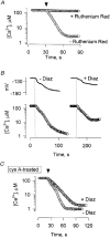
A, Ca2+ concentration within the mitochondrial suspension in the presence and absence of Ruthenium Red (100 nM), an inhibitor of the mitochondrial membrane potential-dependent Ca2+ uniporter. Arrowhead indicates addition of mitochondria (1 mg protein) to a solution containing 150 μm Ca2+. B, time course of mitochondrial membrane potential during Ca2+ uptake in the absence and presence of 300 μm diazoxide. Extramitochondrial Ca2+ and TPP+ (membrane potential) were simultaneously recorded using corresponding ion-selective minielectrodes. C, diazoxide (300 μm) reduced the rate of Ca2+ uptake in mitochondria treated with 2 μm cyclosporin A (cys A), an inhibitor of the mitochondrial permeability transition pore.
Figure 3
Figure 3. Potassium channel openers unload Ca2+ from pre-loaded mitochondria
A, Ca2+ concentration within a mitochondrial suspension in the absence and presence of 30 and 300 μm diazoxide. Inset, effect of 600 μm pinacidil. Arrowheads indicate addition of mitochondria (1 mg protein) to a solution containing 150 μm Ca2+. Arrows indicate addition of the respective potassium channel openers. B, concentration-response curve for the effect of diazoxide on the inhibition of the rate of mitochondrial Ca2+ release. Inset, concentration-response curve for the effect of pinacidil. Curves were fitted using the Hill equation. Data are from 5-10 mitochondrial preparations.
Figure 4
Figure 4. Effect of cyclosporin A on potassium channel opener-induced membrane depolarization and Ca2+ release
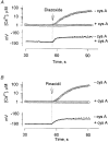
Time course of Ca2+ discharge and membrane depolarization in mitochondria pre-loaded with 150 nmol Ca2+ (mg protein)−1 following addition of 300 μm diazoxide (A) or 600 μm pinacidil (B) (arrows) in the presence (+ cys A) or absence (- cys A) of 2 μm cyclosporin A. Extramitochondrial Ca2+ and TPP+ (membrane potential) were simultaneously recorded using corresponding ion-selective minielectrodes.
Figure 5
Figure 5. Replacement of K+ with mannitol abolishes the effects of potassium channel openers
A, rate of Ca2+ uptake in the absence (control) or presence of 300 μm diazoxide or 600 μm pinacidil in mitochondria incubated in 110 mM KCl or in nominally K+-free solution (Mannitol), where 110 mM KCl was replaced with 220 mM mannitol. B, rate of Ca2+ release in the absence (control) or presence of 300 μm diazoxide or 600 μm pinacidil by mitochondria incubated in 110 mM KCl or nominally K+-free solution (Mannitol). * indicate significant difference from control.
Figure 6
Figure 6. Valinomycin induces release of Ca2+ from mitochondria
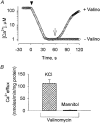
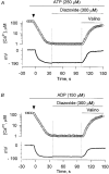
A, Ca2+ concentration within the mitochondrial suspension in the absence and presence of 1 ng valinomycin (mg protein)−1. Arrowhead indicates addition of mitochondria (1 mg protein) to a solution containing 150 μm Ca2+. Arrow indicates addition of valinomycin (Valino). B, rate of Ca2+ release in the presence of 1 ng valinomycin (mg protein)−1 by mitochondria incubated in 110 mM KCl or nominally K+-free solution (Mannitol).
Figure 7
Figure 7. ATP and ADP inhibit diazoxide-induced mitochondrial depolarization and reduction in Ca2+ uptake
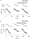
Time course of mitochondrial membrane potential and Ca2+ uptake in the absence (left) and presence of diazoxide (centre), and in the additional presence of a mitoKATP-inhibitory nucleotide (right), ATP (A) or ADP (B). Extramitochondrial Ca2+ and TPP+ (membrane potential) were simultaneously recorded using corresponding ion-selective minielectrodes. Dotted vertical lines indicate addition of mitochondria.
Figure 8
Figure 8. ATP and ADP inhibit diazoxide- but not valinomycin-induced mitochondrial depolarization and Ca2+ release
Time course of Ca2+ discharge and membrane depolarization in mitochondria pre-loaded with 150 nmol Ca2+ (mg protein)−1 following addition of 300 μm diazoxide in the presence of 250 μm ATP (A) or 150 μm ADP (B). Extramitochondrial Ca2+ and TPP+ (membrane potential) were simultaneously recorded using corresponding ion-selective minielectrodes. Arrowheads indicate addition of mitochondria (1 mg protein) to a solution containing 150 μm Ca2+. Experimental conditions as in Fig. 3, except that atractyloside (20 μm) was added to prevent internalization of added nucleotides. The K+ ionophore valinomycin was added as a control.
Figure 9
Figure 9. Effect of nucleotides on diazoxide-induced Ca2+ release from pre-loaded cardiac mitochondria
Relative rate of 300 μm diazoxide-mediated Ca2+ release from pre-loaded mitochondria in the presence of ATP (250 μm), GTP (250 μm), UTP (250 μm), ADP (150 μm), GDP (250 μm) and UDP (250 μm). In each case, the rate of efflux is expressed as a percentage of the rate induced by 25 μm 2,4-dinitrophenol, the mitochondrial uncoupler, determined in the same mitochondrial preparation.
Figure 10
Figure 10. Diazoxide-induced mitochondrial depolarization in intact cardiomyocytes: prevention by the mitoKATP channel blocker 5-HD
A, confocal images of TMRM-loaded neonatal cardiomyocytes in the absence (- Diaz) and presence (+ Diaz) of 300 μm diazoxide. Note the decrease in fluorescence following addition of the potassium channel opener, indicative of opener-induced membrane depolarization. B, confocal images of TMRM-loaded neonatal cardiomyocytes treated with 300 μm 5-HD in the absence (5-HD) and presence of 300 μm diazoxide (+ Diaz) or 1 μg ml−1 FCCP (+ FCCP). Note the absence of a fluorescence change following addition of the potassium channel opener. Horizontal bar, 10 μm. Vertical bar indicates relative fluorescence scale (white indicates highest fluorescence).
Figure 11
Figure 11. Diazoxide-induced decrease in mitochondrial Ca2+ load in intact cardiomyocytes: prevention by the mitoKATP channel blocker 5-HD
A, confocal images of rhod-2 AM-loaded neonatal cardiomyocytes in the absence (- Diaz) and presence (+ Diaz) of 300 μm diazoxide. Note the decrease in fluorescence following addition of the potassium channel opener, indicative of an opener-induced decrease in mitochondrial Ca2+ content. B, confocal images of rhod-2 AM-loaded neonatal cardiomyocytes treated with 300 μm 5-HD in the absence (5-HD) and presence of 300 μm diazoxide (+ Diaz) or 1 μg ml−1 FCCP (+ FCCP). Note the absence of a fluorescence change following addition of the potassium channel opener. Horizontal bar, 10 μm. Vertical bar indicates relative fluorescence scale (white indicates highest fluorescence).

Similar articles

Cited by

References

    1. Alekseev AE, Brady PA, Terzic A. Ligand-insensitive state of cardiac ATP-sensitive K+ channels: Basis for channel opening. Journal of General Physiology. 1998;111:381–394. - PMC - PubMed
    1. Bassani JW, Bassani RA, Bers DM. Ca2+ cycling between sarcoplasmic reticulum and mitochondria in rabbit cardiac myocytes. The Journal of Physiology. 1993;460:603–621. - PMC - PubMed
    1. Beavis AD, Lu Y, Garlid KD. On the regulation of K+ uniport in intact mitochondria by adenine nucleotides and nucleotide analogs. Journal of Biological Chemistry. 1993;268:997–1004. - PubMed
    1. Belyaeva E, Szewczyk A, Mikolaek B, Nalecz M, Wojtczak L. Demonstration of glibenclamide-sensitive K+ fluxes in rat liver mitochondria. Biochemistry and Molecular Biology International. 1993;31:493–500. - PubMed
    1. Bernardi P, Petronilli V. The permeability transition pore as a mitochondrial Ca2+ release channel: A critical appraisal. Journal of Bioenergetics and Biomembranes. 1996;28:131–138. - PubMed

Publication types

MeSH terms

LinkOut - more resources