. 1999 Sep 1;519 Pt 2(Pt 2):427-37.
doi: 10.1111/j.1469-7793.1999.0427m.x.

Activity-dependent modulation of K+ currents at presynaptic terminals of mammalian central synapses

Affiliations

Activity-dependent modulation of K+ currents at presynaptic terminals of mammalian central synapses

J Qian et al. J Physiol. .

Abstract

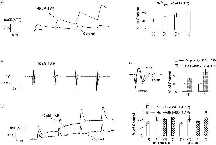
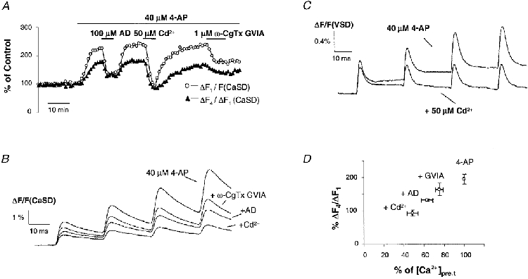
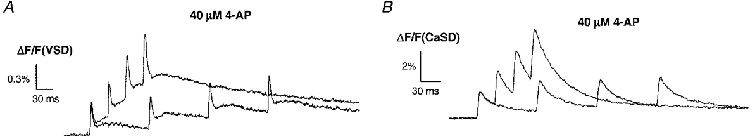
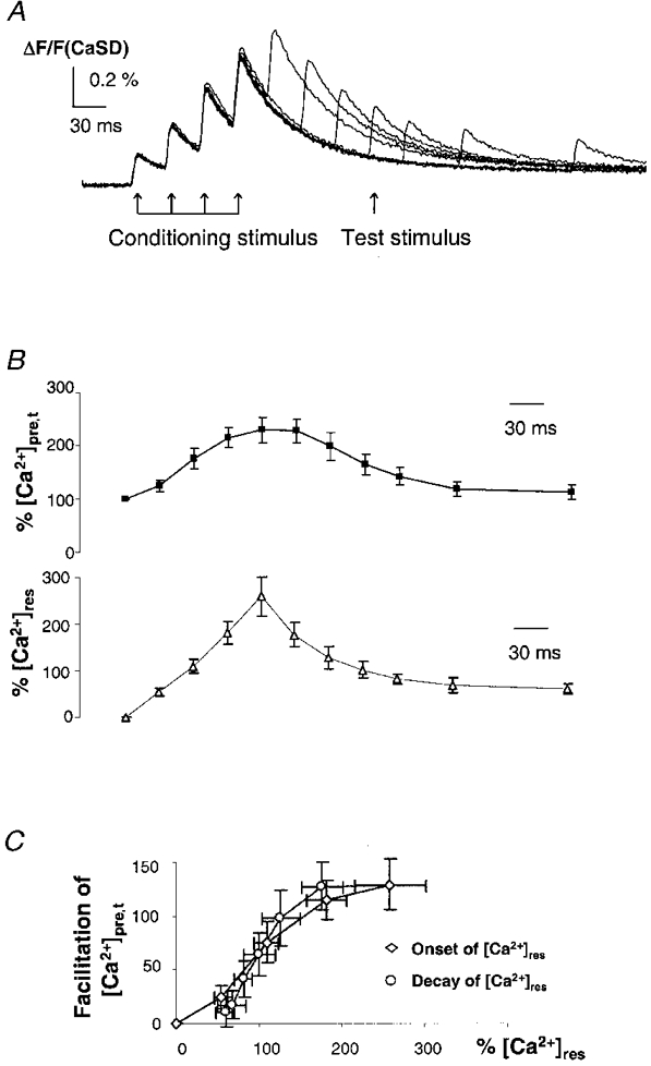
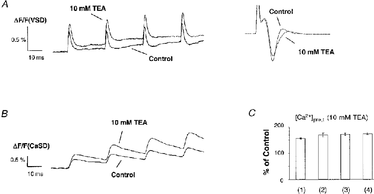
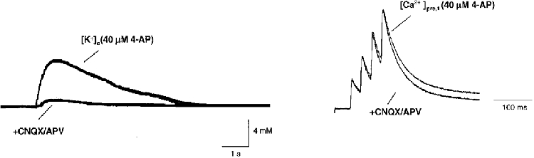
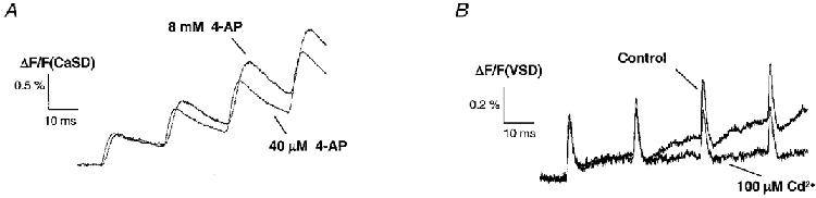
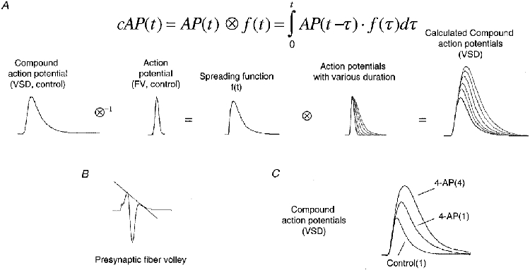
1. The activity-dependent regulation of presynaptic K+ currents at the CA3-CA1 synapse in the rat hippocampus was investigated during a train of evoked afferent action potentials. The waveforms of presynaptic compound action potentials (cAPs) and presynaptic Ca2+ transients ([Ca2+]pre,t) were measured with fluorescent voltage-sensitive and Ca2+-sensitive indicators in rat brain slices. 2. Under control conditions, presynaptic cAPs and the accompanying [Ca2+]pre,t displayed similar amplitudes for each stimulus, suggesting that there was no cumulative change of K+ and Ca2+ currents during the test train. However, when a subgroup of presynaptic K+ channels was blocked by a low concentration of 4-aminopyridine (4-AP, 40 microM), a significant facilitation of the [Ca2+]pre,t was observed. 3. This phenomenon was not due to a direct action of 4-AP on presynaptic Ca2+ channels, but to cumulative suppression of the K+ conductance as indicated by the corresponding change in waveforms of the cAP and presynaptic fibre volley. The observed facilitation was not an artifact by virtue of increased fibre recruitment, nor was it related to the accumulation of extracellular K+; rather, it was dependent on Ca2+ influx and stimulation frequency. The time course of recovery from facilitation was closely related to the decay of the intracellular Ca2+ concentration. 4. The facilitation was not blocked by a saturating concentration of 4-AP (8 mM) but was reduced during the application of the K+ channel blocker tetraethylammonium (TEA, 10 mM), implicating the involvement of TEA-sensitive K+ channels. Such activity-dependent suppression of presynaptic K+ conductance could lead to excessive transmitter release and might explain the hippocampal epileptiform activity that can be induced by application of 4-AP.


Figures

Figure 1
Figure 1. Presynaptic signals under control conditions
Sample traces of presynaptic compound action potentials (cAPs), fibre volleys (FVs) and Ca2+ transients ([Ca2+]pre,t) in response to a train of four stimuli at an interpulse interval of 30 ms under control conditions. A, cAPs measured with the VSD RH 414. In addition to the rapid change of membrane potential that corresponds to the presynaptic action potential, the VSD signal also contains a slow component. B, corresponding presynaptic FVs measured by extracellular field recording. C, [Ca2+]pre,t, measured separately with the Ca2+ indicator furaptra. D, summary data for cAP, FV (n = 12) and [Ca2+]pre,t (n = 18). The responses to each stimulus within the train were normalized to the mean of the last three. There was no significant difference in the amplitudes of FV and [Ca2+]pre,t between stimuli. Amplitude and half-width of the uncorrected cAP in response to the first stimulus were larger than to the rest of the stimuli. This was most probably due to the artifact of the slow component.
Figure 2
Figure 2. 4-AP increases and facilitates both [Ca2+]pre,t and cAP
Sample traces of [Ca2+]pre,t, FVs and cAPs in response to a train of stimuli under control conditions and in 4-AP. Application of 40 μm 4-AP broadened the FV, increased the [Ca2+]pre,t and prolonged repolarization of the VSD signals. In contrast to a similar duration of FVs for each stimulus in control, FVs were progressively broadened for the subsequent stimuli in 4-AP. Consistently, the amplitudes of [Ca2+]pre,t were facilitated in 4-AP. A, on average, the [Ca2+]pre,t was increased to 226 ± 36, 278 ± 40, 356 ± 47 and 437 ± 47 % of control (n = 8) during application of 4-AP for successive stimuli within the train. B, the half-width of FV in response to the first and last stimulus was 162 ± 13 and 248 ± 38 % of control (n = 12) during application of 4-AP, respectively. The amplitude of the FV was not significantly changed (see inset). This argues against a possible recruitment of presynaptic fibres by the subsequent stimuli. C, the slow component of the VSD signal during application of 4-AP was corrected for by subtracting fitted exponential functions (thin line). The chart summarizes amplitude and half-width of VSD signals for the first and last stimulus in the presence of 4-AP. The left-hand side shows raw data of the first and last cAP (n = 12; amplitude, 164 ± 11 and 213 ± 23 %; half-width, 201 ± 18 and 229 ± 30 %); the right-hand side shows corrected data (n = 12; amplitude, 152 ± 14 and 200 ± 24 %; half-width, 179 ± 15 and 236 ± 31 %). There was a significant increase in both amplitude and half-width of the cAP waveform after correction for the slow component (paired two-tailed t test, * P < 0.001).
Figure 3
Figure 3. Facilitation of [Ca2+]pre,t is dependent on Ca2+ influx
A, time course of normalized [Ca2+]pre,t and its facilitation from a typical experiment. The amount of facilitation is presented as the amplitude ratio of the last to the first Ca2+ influx (ΔF4F1). In the presence of 4-AP, application of adenosine (AD), Cd2+ and ω-CgTX GVIA inhibited the [Ca2+]pre,t and reduced the facilitation. B, sample traces of the CaSD signals shown in A. C, VSD signals in the presence of 4-AP and during application of Cd2+. This non-specific blocker of voltage-dependent Ca2+ channels eliminated the progressive increase of cAP waveform, consistent with the Ca2+ dependency observed in the CaSD signal. D, summary data illustrating that the facilitation of [Ca2+]pre,t was dependent on presynaptic Ca2+ influx. In the presence of 4-AP (40 μm), application of ω-CgTX GVIA (1 μm), AD (100 μm) and Cd2+ (30 μm) inhibited [Ca2+]pre,t by 25 ± 4 % (n = 8), 37 ± 6 % (n = 4) and 51 ± 6 % (n = 7), respectively. The corresponding response to the fourth stimulus was reduced to 164 ± 19, 133 ± 3 and 93 ± 9 % of the first.
Figure 4
Figure 4. Facilitation of [Ca2+]pre,t depends on the frequency of stimulation
Sample traces of cAPs (A) and [Ca2+]pre,t (B) evoked by stimulation with different interstimulus intervals in the presence of 4-AP. Facilitation of cAPs was dependent on the stimulation frequency. When stimuli were delivered at an interval of 100 ms, there was no progressive increase in the amplitude of the cAP. The facilitation of the [Ca2+]pre,t shows a similar frequency dependence to the cAP.
Figure 5
Figure 5. Recovery time course of [Ca2+]pre,t facilitation
A, [Ca2+]pre,t from a typical experiment to measure the time course of recovery from facilitation of [Ca2+]pre,t. Following the conditioning stimulation train, a single test stimulus was applied at various intervals. B, summary data to show the time course of [Ca2+]pre,t facilitation and [Ca2+]res (n = 8). The recovery is represented by the [Ca2+]pre,t in response to the test stimulus after the conditioning stimulation, which was measured after subtracting the decay time course of residual Ca2+ traces ([Ca2+]res). By visual inspection, the recovery time course for the facilitation appears similar to the decay of the [Ca2+]res. This suggests that the suppression of K+ currents is related to the [Ca2+]res. C, relationship between facilitation of [Ca2+]pre,t and normalized [Ca2+]res during conditioning stimulation and testing. The onset and decay phases of [Ca2+]res exhibit a slightly different relationship with [Ca2+]pre,t. This difference may reflect a small amount of cumulative voltage-dependent modulation during the conditioning stimulation.
Figure 6
Figure 6. Effects of TEA on the cAP and [Ca2+]pre,t
Sample traces of cAPs and [Ca2+]pre,t under control conditions and in the presence of 10 mM TEA. A, in contrast to application of 4-AP, TEA did not evoke a progressive increase in the amplitude of VSD signals. No difference was detected in the waveform of the FV among stimuli within the test train. The inset shows superimposed presynaptic FVs. TEA not only increased the duration but also reduced the amplitude of the FV compared with control, suggesting a decreased excitability of fibres in the presence of high concentrations of TEA. B, [Ca2+]pre,t in control and in the presence of 10 mM TEA. Consistent with the observed cAP, there was much less facilitation of the [Ca2+]pre,t for subsequent stimuli within the test train. C, summary data for the [Ca2+]pre,t in the presence of TEA. On average, 10 mM TEA increased the [Ca2+]pre,t to 156 ± 3, 168 ± 7, 171 ± 5 and 174 ± 4 % of control (n = 4) for successive stimuli within the test train. The amount of [Ca2+]pre,t induced by 10 mM TEA is probably an underestimate, due to the reduced amount of fibres activated. There was no significant facilitation of [Ca2+]pre,t as compared with application of 4-AP.
Figure 7
Figure 7. Modulation of K+ conductance is not related to [K+]o
[K+]o and [Ca2+]pre,t in the presence of 4-AP and during application of the glutamate receptor antagonists CNQX and D-APV. Blockade of postsynaptic responses with 10 μm CNQX and 25 μm D-APV did not eliminate the facilitation of [Ca2+]pre,t while this manipulation greatly reduced the stimulation-evoked increase of [K+]o. This suggests that the observed activity-dependent modulation of K+ conductance was not related to [K+]o.
Figure 8
Figure 8. Facilitation of [Ca2+]pre,t is insensitive to high concentrations of 4-AP
A, [Ca2+]pre,t in the presence of 8 mM 4-AP. This concentration is supposed to block fast inactivating (A-type) K+ currents; however, it did not eliminate the facilitation of [Ca2+]pre,t, suggesting that A-type K+ channels were not involved. High concentrations of 4-AP reduced the amplitude of FVs (not shown). Therefore, the trace of [Ca2+]pre,t shown here is an underestimate of presynaptic Ca2+ influx as compared with application of 40 μm 4-AP. B, application of Cd2+ (100 μm), abolished the afterhyperpolarization, suggesting the existence of presynaptic Ca2+-sensitive K+ currents.
Figure 9
Figure 9. Interpretation of cAPs measured with voltage-sensitive dyes
Due to the nature of the optical recording technique, a cAP measured with the VSD is actually a convolution of the waveform of the true action potential (AP) with a spreading function, f(t), of the AP within the optical recording area. A, to estimate the spreading function, a cAP measured under control conditions was de-convoluted with the waveform of an AP, obtained from a corrected presynaptic fibre volley in control (see B). Then, cAPs for APs of various durations were obtained by convoluting the waveform of APs with the estimated spreading function. Gaussian functions were used to simulate the waveform of action potential repolarization. B, mean waveform of presynaptic FVs under control conditions. The dotted line indicates the correction for the field effect. C, mean waveform of cAPs after correction for the slow component under control conditions and during application of 4-AP. These cAPs measured with the VSD show patterns similar to the calculated waveforms of cAPs.

Similar articles

Cited by

References

    1. Aldrich RW, Getting PA, Thompson SH. Mechanisms of frequency-dependent broadening of molluscan neurone soma spikes. The Journal of Physiology. 1979;291:531–544. - PMC - PubMed
    1. Augustine GJ. Regulation of transmitter release at the squid giant synapse by presynaptic delayed rectifier potassium current. The Journal of Physiology. 1990;431:343–364. - PMC - PubMed
    1. Bielefeldt K, Rotter JI, Jackson MB. Three potassium channels in rat posterior pituitary nerve endings. The Journal of Physiology. 1992;458:41–67. - PMC - PubMed
    1. Brigant DC, Mallart A. Presynaptic currents in mouse motor endings. The Journal of Physiology. 1982;333:619–639. - PMC - PubMed
    1. Chen QX, Wong RKS. Intracellular Ca2+ suppressed a transient potassium current in hippocampal neurons. Journal of Neuroscience. 1991;11:337–343. - PMC - PubMed

Publication types

MeSH terms

LinkOut - more resources