. 1999 Sep 15;519 Pt 3(Pt 3):669-78.
doi: 10.1111/j.1469-7793.1999.0669n.x.

Kinetic differences at the single molecule level account for the functional diversity of rabbit cardiac myosin isoforms

Affiliations

Kinetic differences at the single molecule level account for the functional diversity of rabbit cardiac myosin isoforms

K A Palmiter et al. J Physiol. .

Abstract

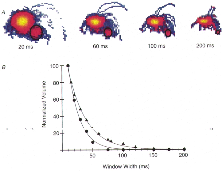
1. Cardiac V3 myosin generates slower actin filament velocities and higher average isometric forces (in an in vitro motility assay) when compared with the V1 isoform. 2. To account for differences in V1 and V3 force and motion generation at the molecular level, we characterized the mechanics and kinetics of single V1 and V3 myosin molecules using a dual laser trap setup. 3. No differences in either unitary displacement (approximately 7 nm) or force (approximately 0.8 pN) were observed between isoforms; however, the duration of unitary displacement events was significantly longer for the V3 isoform at MgATP concentrations > 10 microM. 4. Our results were interpreted on the basis of a cross-bridge model in which displacement event durations were determined by the rates of MgADP release from, and MgATP binding to, myosin. 5. We propose that the release rate of MgADP from V3 myosin is half that of V1 myosin without any difference in their rates of MgATP binding; thus, kinetic differences between the two cardiac myosin isoforms are sufficient to account for their functional diversity.


Figures

Figure 1
Figure 1. Determination of unitary step displacement (d) and force (F) in V1 and V3 cardiac myosin
A, 5 s of time-series displacement data obtained with V1 myosin (upper trace) and V3 myosin (lower trace) at 0.005 mM MgATP. Arrows bracket discrete displacement events. B, corresponding raw and baseline-subtracted (- Baseline) MV displacement histograms for the complete (≈60 s) time-series data shown in Fig. 1A. The MV histograms were generated using a 20 ms window. For a complete description of MV analysis see Methods. The letters ‘B’ and ‘e’ identify the baseline and event populations, respectively. MV histograms are presented 2-dimensionally with the third dimension, i.e. counts, colour mapped. A colour bar (log scale) that applies to all histograms is illustrated, with minimum and maximum counts of -222 and 1268 and -270 and 2572 for the V1 and V3 histograms, respectively. For these data, the event populations for the V1 and V3 were fitted at 4.9 ± 0.1 nm (mean ±s.d.) and 5.4 ± 0.1 nm, respectively. See Table 1 for compilation of all d values for the various MgATP concentrations studied. Occasionally, events in the negative direction are observed and are noted in the V1 MV histogram by an arrow. C, 5 s of time-series force data obtained with V1 myosin (2 upper traces) and V3 myosin (2 lower traces) at 0.005 mM MgATP. For each isoform, the upper time-series trace is the AOM correction signal, which is calibrated to force. The lower trace is the residual displacement trace in feedback mode, i.e. the motion of the bead while under feedback control. Arrows bracket discrete unitary force events, where the reduction in variance is most apparent on the residual displacement trace. D, corresponding raw and baseline-subtracted (- Baseline) MV force histograms generated using a 20 ms window from the complete time-series force data shown in Fig. 1C. The colour bar (log scale) minimum and maximums were -148 and 769 and -178 and 1142 for the V1 and V3 isoforms, respectively. The event populations in the V1 and V3 histograms were fitted at 0.7 ± 0.02 and 1.2 ± 0.02 pN, respectively.
Figure 2
Figure 2. Determination of ton
A, representative MV histogram illustrating the exponential decay in the event population volume (encircled by black ellipse) as window width is increased. B, representative volume versus window width curves generated from data collected with V1 and V3 myosin at 0.010 mM MgATP. To determine average ton a gallery of MV histograms generated at different time window widths (10, 15, 20, 30, 40, 50, 60, 75, 80, 100, 120, 150, 175 and 200 ms) was created. The number of counts in the event populations of the various MV histograms was recorded. As illustrated in Fig. 2A the volume of counts decays exponentially as time window widths increase. For illustrative purposes, the data were normalized to the maximum number of counts obtained at the 10 ms window width. As with all data, the normalized data were fitted to the equation: V =N tone-W/ton. N is the estimated number of events within the record and W is the window width. The V1 plot (•) was fitted with ton= 18 ± 0.01 ms (mean ± s.e.) and N = 81± 4, r2= 0.997 whereas the V3 plot (▴) was fitted with ton= 31 ± 0.01 ms with an N = 320± 13, r2= 0.999. Estimates of N vary with the total record length. The representative V1 and V3 data sets were chosen because their average ton values were similar to the mean ton calculated for all V1 and V3 data, respectively, at 0.01 mM MgATP.
Figure 3
Figure 3. Representative displacement time-series data traces (5 s) obtained with V1 cardiac myosin at various MgATP concentrations
Arrows bracket discrete unitary displacement events (at 1 mM MgATP one arrow identifies each event). As shown there are obvious differences in the durations of unitary events at different MgATP concentrations. At 1 mM MgATP events become too fast to visually identify and thus the MV analysis becomes essential for estimating d and ton. The estimated d and ton for the V1 and V3 myosin for each [MgATP] range are given in Table 1.
Figure 4
Figure 4. Kinetic model to account for functional diversity between cardiac myosin isoforms
A, kinetic model of ton. The duration of strong actin-myosin (AM) binding can be modelled as two discrete kinetic steps, t-ADP and t+ATP, which correspond to biochemical transitions in the cross-bridge cycle. At physiological MgATP concentrations ton will be predominantly determined by t-ADP, which is related to the rate of ADP release from the active site. Thus the rigor, AM state, will be very short lived. However, as MgATP concentrations decrease, more of the total ton duration will be accounted for by the time spent waiting for ATP rebinding (t+ATP) and the rigor AM state is prolonged. B, relationship between ton and [MgATP]. V1 (•) and V3 (▴) ton data are expressed as the mean ±s.e.m. at the indicated [MgATP] (see Table 1). tonversus[MgATP] data were fitted by least squares regression analysis using TableCurve 2D software (SPSS Inc., Chicago, IL, USA) to eqn (2) as described in the text.

References

    1. Alousi AA, Grant AM, Etzler JR, Cofer BR, van der Bel-Kahn J, Melvin D. Reduced cardiac myofibrillar Mg-ATPase activity without changes in myosin isoenzymes in patients with end-stage failure. Molecular and Cellular Biochemistry. 1990;96:79–88. - PubMed
    1. Alpert NR, Mulieri LA. Increased myothermal economy of isometric force generation in compensated cardiac hypertrophy induced by pulmonary artery constriction in the rabbit. Circulation Research. 1982;50:491–500. - PubMed
    1. Cappelli V, Bottinelli R, Poggesi C, Moggio R, Reggiani C. Shortening velocity and myosin and myofibrillar ATPase activity related to myosin isoenzyme composition during postnatal development in rat myocardium. Circulation Research. 1989;65:446–457. - PubMed
    1. Dantzig JA, Goldman YE, Millar NC, Lacktis J, Homsher E. Reversal of the cross-bridge force-generating transition by photogeneration of phosphate in rabbit psoas muscle fibres. The Journal of Physiology. 1992;451:247–278. - PMC - PubMed
    1. Dupuis DE, Guilford WH, Wu J, Warshaw DM. Actin filament mechanics in the laser trap. Journal of Muscle Research and Cell Motility. 1997;18:17–30. - PubMed

Publication types

LinkOut - more resources