. 1999 Aug;97(4):626-33.
doi: 10.1046/j.1365-2567.1999.00818.x.

Enhanced antigen-presenting activity and tumour necrosis factor-alpha-independent activation of dendritic cells following treatment with Mycobacterium bovis bacillus Calmette-Guérin

Affiliations

Enhanced antigen-presenting activity and tumour necrosis factor-alpha-independent activation of dendritic cells following treatment with Mycobacterium bovis bacillus Calmette-Guérin

K D Kim et al. Immunology. 1999 Aug.

Abstract

Dendritic cells (DCs) are most potent among the antigen-presenting cells and are believed to be crucial for the initiation of a primary T-cell response to foreign antigens. Mycobacterial infection within macrophages is controlled by cell-mediated immunity. To elucidate the stimulation of immune response by Mycobacterium bovis bacillus Calmette-Guérin (BCG), we purified DCs from precursor cells in human peripheral blood mononuclear cells (PBMC) by culturing them with granulocyte-macrophage colony-stimulating factor (GM-CSF) and interleukin-4 (IL-4) and characterized their surface antigen expression. The interaction of cultured DCs with BCG resulted in increased surface expression of several DC-related marker antigens. BCG also induced reduction of endocytosis, enhancement of CD83 expression as well as B7 costimulatory molecules and IL-12 production, suggesting that BCG treatment directly induces DCs to mature. BCG-treated DCs were much more potent antigen-presenting cells in allogeneic immune response than untreated DCs. Moreover, while the neutralization of tumour necrosis factor-alpha (TNF-alpha) significantly blocked the DC maturation induced by lipopolysaccharide (LPS), it could not inhibit the induction of DC maturation by the BCG treatment, indicating that TNF-alpha production plays a minor role in the BCG-induced DC maturation. However, the neutralization of TNF-alpha resulted in decreased IL-12 production by activated DCs. These results suggest that infection with BCG might evoke direct activation and maturation of DC and the general immune stimulant effect of BCG might be related with the activation of DCs.


Figures
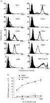
Figure 1
Figure 1
(a) Phenotype of dendritic cells. DCs were generated as described in Materials and Methods. They were harvested after a culture of 6 days, washed and stained with respective mAbs. The surface antigen expression was measured with a flow cytometer (Beckton Dickinson) and analysed with FACScan® and LYSYS® software. The filled histogram shows patterns obtained with isotype-matched control antibodies. (b) DCs derived from culture of human PBMC function as potent APC. CD3-positive T cells (5×10⁴) (>70%) purified from PBMC using a human T-cell enrichment column were incubated with various numbers of irradiated (6000 rad) allogeneic whole PBMC, monocytes or DCs, respectively. The proliferation of T cells in triplicate was determined after 6 days by pulsing with 1 μCi of [3H]thymidine for the last 18 hr and counting with liquid scintillation counter. This is a representative experiment of three independent experiments with similar results.
Figure 2
Figure 2
BCG treatment upregulates surface antigen expression on DCs. DCs generated from plastic-adherent cells as described in Materials and Methods were incubated without BCG or with 1·5×105 CFU of BCG for 24 hr in cytokine-free culture medium. Cells were stained and analysed as described in Fig. 1. Numbers indicate percentage increase of mean fluorescence intensity (MFI). The MFI values of cells stained with isotype-matched control antibodies were 3·8 for FITC-control and 3·5 for PE-control. This is a representative experiment of four independent experiments with relatively similar percentage increases.
Figure 3
Figure 3
Enhancement of allogeneic T-cell stimulation by BCG-treated DCs. DCs were treated with or without BCG (1·5×105 CFU) for 24 hr, washed thoroughly and added to either purified allogeneic T cells (circles) or autologous BCG-primed T cells from vaccinated donor (triangles). On day 6, proliferation was measured by thymidine uptake for 18 hr.
Figure 4
Figure 4
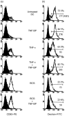
BCG induces maturation of DCs. DCs (1×10⁵) were treated either with 50 ng/ml of TNF-α or 5 μg/ml of soluble TNF-binding protein, with live BCG (1·5×105 CFU) or with both of them for 24 hr in culture media without addition of cytokines. (a,b) DCs were stained with isotype control antibody or anti-CD83-PE and CD83 expression was analysed using flow cytometer (a). The endocytic activity of DCs was determined by flow cytometer after treatment with FITC-dextran (b). The control endocytic activity was determined after FITC-dextran treatment at 4°. Numbers indicate the percentage and mean fluorescence intensity of FITC-positive cells exhibiting endocytosis of immature DCs. TNF-binding protein (TNF-BP); mean fluorescence intensity (MFI).
Figure 5
Figure 5
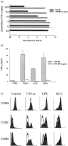
The neutralization of TNF-α activity does not affect BCG-induced DC maturation. (a) Determination of neutralizing activity of TNF-binding protein. TNF-α was added to 1×10⁴ Wehi-164 cells in 96-well culture plate in the absence or presence of 5 μg/ml of TNF-binding protein. The cytotoxic activity of TNF-α was measured by MTS/PMS staining method described in Materials and Methods. (b) Activated DCs produce TNF-α, which can be neutralized by an addition of TNF-binding protein. DCs were treated with TNF-α (50 ng/ml), LPS (200 ng/ml) or BCG (1·5×105 CFU/ml) in the absence or presence of 5 μg/ml TNF-binding protein. After 24 hr, TNF-α content of culture supernatant was assayed by a standard bioassay using Wehi-164 cells. (c) The neutralization of TNF-α activity affects DC maturation induced by LPS but not by BCG. DCs were treated with stimuli in the absence (shaded histogram) or the presence (open histogram) of TNF-binding protein for 24 hr. Cells were harvested and incubated with antibodies against CD80, CD83 and CD86. The expression of surface antigens was analysed using a flow cytometer.
Figure 6
Figure 6
Cultured DCs produce significant levels of bioactive IL-12 following stimulation with BCG. DCs (1×10⁵/ml) cultured in GM-CSF and IL-4 were exposed to the indicated stimuli as described in Fig. 4 and the bioactive p70 heterodimer was measured after 40 hr by an IL-12 enzyme-linked immunosorbent assay (ELISA) kit according to the manufacturer’s instruction. Experiments were performed independently in duplicate with similar results.

References

    1. Crowley M, Inaba K, Steinman RM. Dendritic cells are the principal cells in mouse spleen bearing immunogenic fragments of foreign proteins. J Exp Med. 1990;172:383. - PMC - PubMed
    1. Steinman RM. The dendritic cell system and its role in immunogenicity. Ann Rev Immunol. 1991;9:272. - PubMed
    1. Cella M, Sallusto F, Lanzavecchia A. Origin, maturation and antigen presenting function of dendritic cells. Curr Opin Immunol. 1997;9:10. - PubMed
    1. Freudenthal PS, Steinman RM. The distinct surface of human blood dendritic cells, as observed after an improved isolation method. Proc Natl Acad Sci USA. 1990;87:7698. - PMC - PubMed
    1. O’doherty U, Steinman RM, Peng M, et al. Dendritic cells freshly isolated from human blood express CD4 and mature into typical immunostimulatory dendritic cells after culture in monocyte-conditioned medium. J Exp Med. 1993;178:1067. - PMC - PubMed

Publication types

MeSH terms

LinkOut - more resources