. 1999 Aug 23;146(4):723-30.
doi: 10.1083/jcb.146.4.723.

The ribosome regulates the GTPase of the beta-subunit of the signal recognition particle receptor

Affiliations

The ribosome regulates the GTPase of the beta-subunit of the signal recognition particle receptor

G Bacher et al. J Cell Biol. .

Abstract

Protein targeting to the membrane of the ER is regulated by three GTPases, the 54-kD subunit of the signal recognition particle (SRP) and the alpha- and beta-subunit of the SRP receptor (SR). Here, we report on the GTPase cycle of the beta-subunits of the SR (SRbeta). We found that SRbeta binds GTP with high affinity and interacts with ribosomes in the GTP-bound state. Subsequently, the ribosome increases the GTPase activity of SRbeta and thus functions as a GTPase activating protein for SRbeta. Furthermore, the interaction between SRbeta and the ribosome leads to a reduction in the affinity of SRbeta for guanine nucleotides. We propose that SRbeta regulates the interaction of SR with the ribosome and thereby allows SRalpha to scan membrane-bound ribosomes for the presence of SRP. Interaction between SRP and SRalpha then leads to release of the signal sequence from SRP and insertion into the translocon. GTP hydrolysis then results in dissociation of SR from the ribosome, and SRP from the SR.


Figures

Figure 1
Figure 1
Binding of GTP to SR. A, Purified SR reconstituted in liposomes analyzed by SDS-PAGE followed by silver staining (left). α[32P]GTP was incubated with SR liposomes (25 nM) and cross-linked to SR by UV irradiation. The sample was subsequently analyzed by SDS-PAGE and PhosphorImaging (right). An unidentified protein of ∼50 kD, found in various amounts in SR preparations, was also labeled with α[32P]GTP and is marked by an asterisk. B, Competition of α[32P]GTP cross-linking to SR (25 nM) reconstituted into liposomes by increasing concentrations of GTP. Radiolabeled SRα was quantified using a PhosphorImager and plotted against the concentration of GTP. Curve connection data points and the apparent Kd of SRα for GTP (14 μM) were calculated by a nonlinear regression program. C, Cross-linking of increasing concentrations of α[32P]GTP to SR (25 nM) reconstituted into liposomes. Radiolabeled SRβ was quantified using a PhosphorImager and plotted against the concentration of α[32P]GTP. The apparent Kd of SRβ for GTP was 20 nM.
Figure 1
Figure 1
Binding of GTP to SR. A, Purified SR reconstituted in liposomes analyzed by SDS-PAGE followed by silver staining (left). α[32P]GTP was incubated with SR liposomes (25 nM) and cross-linked to SR by UV irradiation. The sample was subsequently analyzed by SDS-PAGE and PhosphorImaging (right). An unidentified protein of ∼50 kD, found in various amounts in SR preparations, was also labeled with α[32P]GTP and is marked by an asterisk. B, Competition of α[32P]GTP cross-linking to SR (25 nM) reconstituted into liposomes by increasing concentrations of GTP. Radiolabeled SRα was quantified using a PhosphorImager and plotted against the concentration of GTP. Curve connection data points and the apparent Kd of SRα for GTP (14 μM) were calculated by a nonlinear regression program. C, Cross-linking of increasing concentrations of α[32P]GTP to SR (25 nM) reconstituted into liposomes. Radiolabeled SRβ was quantified using a PhosphorImager and plotted against the concentration of α[32P]GTP. The apparent Kd of SRβ for GTP was 20 nM.
Figure 4
Figure 4
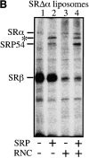
GTP binding to SRβ in the presence of RNC. A, SR liposomes were treated with 2 ng/ml of trypsin (SRΔα liposomes) and analyzed by SDS-PAGE, followed by Western blotting using antibodies raised against SRα and SRβ. B, Cross-linking of radiolabeled GTP to the SRβ in the presence of RNC. Liposomes containing 25 nM SRβ (SRΔα-liposomes) were incubated with 0.3 μM α[32P]GTP in the presence or absence of RNC (8.4 OD260/ml) and/or 20 nM SRP. Samples were UV irradiated and subsequently analyzed by SDS-PAGE and PhosphorImaging. An unidentified protein of ∼65 kD in SRP preparations was also labeled with α[32P]GTP and is marked by an asterisk.
Figure 4
Figure 4
GTP binding to SRβ in the presence of RNC. A, SR liposomes were treated with 2 ng/ml of trypsin (SRΔα liposomes) and analyzed by SDS-PAGE, followed by Western blotting using antibodies raised against SRα and SRβ. B, Cross-linking of radiolabeled GTP to the SRβ in the presence of RNC. Liposomes containing 25 nM SRβ (SRΔα-liposomes) were incubated with 0.3 μM α[32P]GTP in the presence or absence of RNC (8.4 OD260/ml) and/or 20 nM SRP. Samples were UV irradiated and subsequently analyzed by SDS-PAGE and PhosphorImaging. An unidentified protein of ∼65 kD in SRP preparations was also labeled with α[32P]GTP and is marked by an asterisk.
Figure 2
Figure 2
GTP binding to SR in the presence of TRAM and the Sec61p complex. Purified SR, purified TRAM protein, and purified Sec61p complex reconstituted in liposomes were analyzed by SDS-PAGE (10–15% acrylamide gel), followed by silver staining (left). Liposomes containing SR (25 nM), Sec61p complex (∼200 nM), and TRAM protein (∼200 nM) were incubated with 0.3 μM α[32P]GTP. Samples were UV irradiated and subsequently analyzed by SDS-PAGE (12.5% acrylamide gel) and PhosphorImaging. An unidentified protein of ∼50 kD, found in various amounts in SR preparations, was also labeled with α[32P]GTP and is marked by an asterisk.
Figure 3
Figure 3
GTP binding to SR in the presence of RNC. A, Cross-linking of radiolabeled GTP to SR in the presence or absence of RNC and/or SRP. α[32P]GTP (0.3 μM) was incubated with liposomes containing purified SR (25 nM) in the presence or absence of RNC (5.6 OD260/ml) and/or 20 nM SRP. Samples were UV irradiated and subsequently analyzed by SDS-PAGE and PhosphorImaging. An unidentified protein of ∼50 kD, found in various amounts in SR preparations, was also labeled with α[32P]GTP and is marked by an asterisk. B, Cross-linking of increasing concentrations of α[32P]GTP (500 Ci/mmol) to SR (25 nM) in the presence of RNC (8.4 OD260/ml). Radiolabeled SRβ was quantified using the PhosphorImager and plotted against the concentration of α[32P]GTP. The apparent Kd of SRβ for GTP in the presence of RNC was 1 μM. GTP cross-linking assay was performed at low temperature (0°C) to reduce GTP hydrolysis. α[32P]GDP was <1% of α[32P]GTP in the assay, as determined by thin-layer chromatography.
Figure 3
Figure 3
GTP binding to SR in the presence of RNC. A, Cross-linking of radiolabeled GTP to SR in the presence or absence of RNC and/or SRP. α[32P]GTP (0.3 μM) was incubated with liposomes containing purified SR (25 nM) in the presence or absence of RNC (5.6 OD260/ml) and/or 20 nM SRP. Samples were UV irradiated and subsequently analyzed by SDS-PAGE and PhosphorImaging. An unidentified protein of ∼50 kD, found in various amounts in SR preparations, was also labeled with α[32P]GTP and is marked by an asterisk. B, Cross-linking of increasing concentrations of α[32P]GTP (500 Ci/mmol) to SR (25 nM) in the presence of RNC (8.4 OD260/ml). Radiolabeled SRβ was quantified using the PhosphorImager and plotted against the concentration of α[32P]GTP. The apparent Kd of SRβ for GTP in the presence of RNC was 1 μM. GTP cross-linking assay was performed at low temperature (0°C) to reduce GTP hydrolysis. α[32P]GDP was <1% of α[32P]GTP in the assay, as determined by thin-layer chromatography.
Figure 5
Figure 5
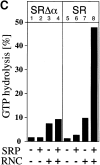
GTP hydrolysis by SR and SRβ in the presence of RNC. A, Hydrolysis of 0.5 μM α[32P]GTP in the presence of different combinations of SR liposomes (40 nM SR), RNC (8.4 OD260/ml), and SRP (50 nM). GTP hydrolysis was stopped by spotting aliquots at different time points onto polyethyleneimine cellulose thin-layer plates. α[32P]GDP was resolved from α[32P]GTP and the amount of α[32P]GDP and α[32P]GTP analyzed by PhosphorImaging. The amount of GTP hydrolyzed was plotted against the incubation time. B, GTP hydrolysis by SR (25 nM) in the presence of different concentrations of RNC. GTP hydrolysis was stopped after 10 min and the amount of α[32P]GDP/GTP analyzed by PhosphorImaging. GTP hydrolysis was plotted against RNC concentrations. Background hydrolysis of GTP by RNC was subtracted. C, GTP hydrolysis by SRβ in the presence of RNC. SRΔα liposomes (40 nM, lanes 1–4) or SR liposomes (lanes 5–8) were incubated with 0.5 μM α[32P]GTP in the presence or absence of 50 nM SRP and/or RNC (8.4 OD260/ml). GTP hydrolysis was stopped after 40 min and the amount of α[32P]GDP/GTP analyzed by PhosphorImaging. Background hydrolysis of GTP by RNC or RNC and SRP, respectively, was subtracted.
Figure 5
Figure 5
GTP hydrolysis by SR and SRβ in the presence of RNC. A, Hydrolysis of 0.5 μM α[32P]GTP in the presence of different combinations of SR liposomes (40 nM SR), RNC (8.4 OD260/ml), and SRP (50 nM). GTP hydrolysis was stopped by spotting aliquots at different time points onto polyethyleneimine cellulose thin-layer plates. α[32P]GDP was resolved from α[32P]GTP and the amount of α[32P]GDP and α[32P]GTP analyzed by PhosphorImaging. The amount of GTP hydrolyzed was plotted against the incubation time. B, GTP hydrolysis by SR (25 nM) in the presence of different concentrations of RNC. GTP hydrolysis was stopped after 10 min and the amount of α[32P]GDP/GTP analyzed by PhosphorImaging. GTP hydrolysis was plotted against RNC concentrations. Background hydrolysis of GTP by RNC was subtracted. C, GTP hydrolysis by SRβ in the presence of RNC. SRΔα liposomes (40 nM, lanes 1–4) or SR liposomes (lanes 5–8) were incubated with 0.5 μM α[32P]GTP in the presence or absence of 50 nM SRP and/or RNC (8.4 OD260/ml). GTP hydrolysis was stopped after 40 min and the amount of α[32P]GDP/GTP analyzed by PhosphorImaging. Background hydrolysis of GTP by RNC or RNC and SRP, respectively, was subtracted.
Figure 6
Figure 6
Interaction of RNC with SRβ in the presence of guanine nucleotides. Liposomes lacking SR (lane 1), SR liposomes (50 nM SR; lanes 2–5), or SRΔα liposomes (50 nM SRβ; lanes 6–9) were incubated with 2.8 OD260/ml RNCs containing 35S-labeled PPL86 and guanine nucleotides in the presence or absence of 50 nM SRP. The liposomes were floated and recovered in the top fraction. Liposome-bound RNCs containing 35S-labeled PPL86 were then quantified by scintillation counting. Bars indicate mean values of three independent experiments with SD.
Figure 7
Figure 7
Model depicting GTP-dependent steps in SRP/SR-mediated targeting of nascent proteins to the ER membrane. We have shown here that SRβ in its GTP-bound form contacts ribosomes. This interaction stimulates GTPase activity of SRβ. SRβ is proposed to allow SRα to scan the ribosome for the presence of SRP.

Similar articles

Cited by

References

    1. Bacher G., Lütcke H., Jungnickel B., Rapoport T.A., Dobberstein B. Regulation by the ribosome of the GTPase of the signal recognition particle during protein targeting. Nature. 1996;381:248–251. - PubMed
    1. Beckmann R., Bubeck D., Grassucci R., Penczek P., Verschoor A., Blobel G., Frank J. Alignment of conduits for the nascent polypeptide chain in the ribosome–Sec61 complex. Nature. 1997;278:2123–2126. - PubMed
    1. Bernstein H.D., Poritz M.A., Strub K., Hoben P.J., Brenner S., Walter P. Model for signal sequence recognition from amino-acid sequence of 54K subunit of signal recognition particle. Nature. 1989;340:482–486. - PubMed
    1. Borgese D., Mok W., Kreibich G., Sabatini D.D. Ribosome–membrane interaction. In vitro binding of ribosomes to microsomal membranes. J. Mol. Biol. 1974;88:559–580. - PubMed
    1. Bourne H.R., Sanders D.A., McCormick F. The GTPase superfamilyconserved structure and molecular mechanism. Nature. 1991;349:117–127. - PubMed

Publication types

MeSH terms