. 1999 Aug 23;146(4):843-54.
doi: 10.1083/jcb.146.4.843.

Analysis of CD44-containing lipid rafts: Recruitment of annexin II and stabilization by the actin cytoskeleton

Affiliations

Analysis of CD44-containing lipid rafts: Recruitment of annexin II and stabilization by the actin cytoskeleton

S Oliferenko et al. J Cell Biol. .

Abstract

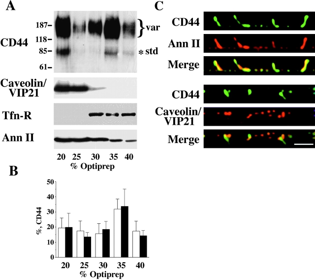
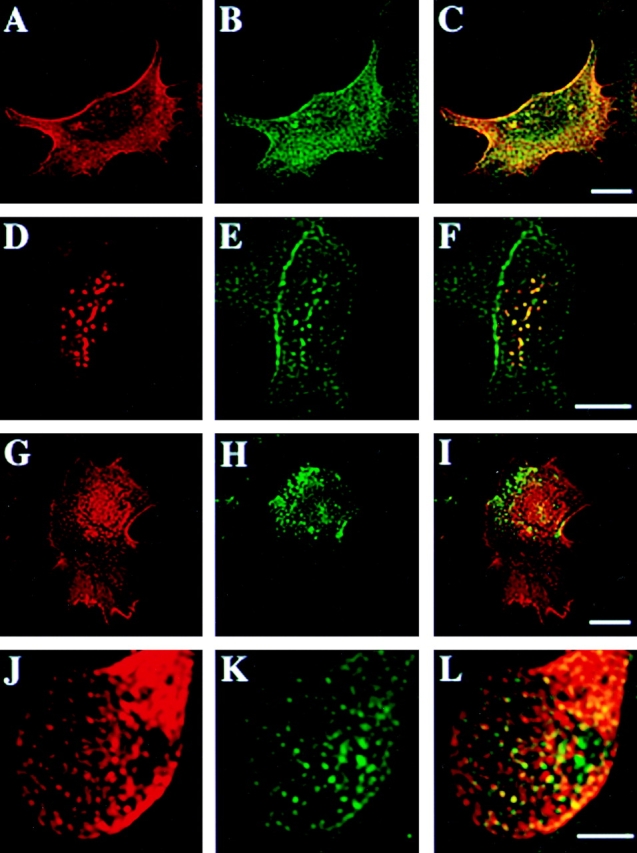
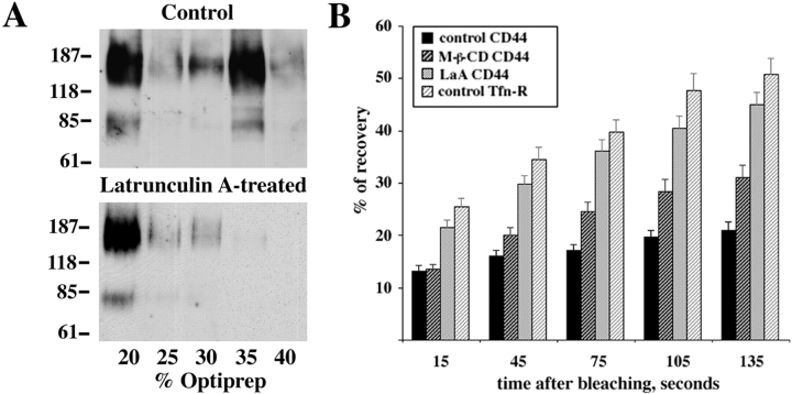
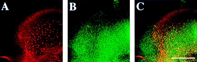
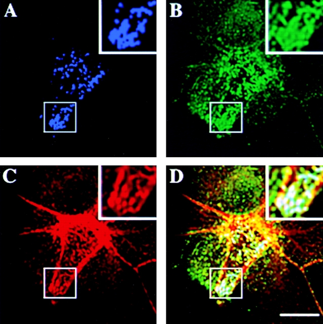
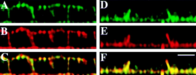
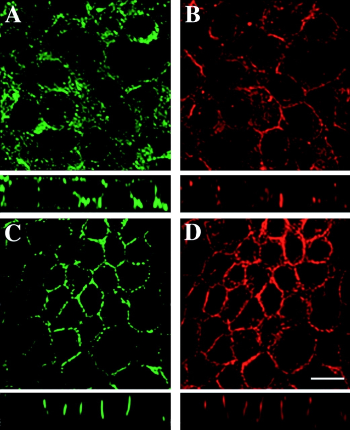
CD44, the major cell surface receptor for hyaluronic acid (HA), was shown to localize to detergent-resistant cholesterol-rich microdomains, called lipid rafts, in fibroblasts and blood cells. Here, we have investigated the molecular environment of CD44 within the plane of the basolateral membrane of polarized mammary epithelial cells. We show that CD44 partitions into lipid rafts that contain annexin II at their cytoplasmic face. Both CD44 and annexin II were released from these lipid rafts by sequestration of plasma membrane cholesterol. Partition of annexin II and CD44 to the same type of lipid rafts was demonstrated by cross-linking experiments in living cells. First, when CD44 was clustered at the cell surface by anti-CD44 antibodies, annexin II was recruited into the cytoplasmic leaflet of CD44 clusters. Second, the formation of intracellular, submembranous annexin II-p11 aggregates caused by expression of a trans-dominant mutant of annexin II resulted in coclustering of CD44. Moreover, a frequent redirection of actin bundles to these clusters was observed. These basolateral CD44/annexin II-lipid raft complexes were stabilized by addition of GTPgammaS or phalloidin in a semipermeabilized and cholesterol-depleted cell system. The low lateral mobility of CD44 in the plasma membrane, as assessed with fluorescent recovery after photobleaching (FRAP), was dependent on the presence of plasma membrane cholesterol and an intact actin cytoskeleton. Disruption of the actin cytoskeleton dramatically increased the fraction of CD44 which could be recovered from the light detergent-insoluble membrane fraction. Taken together, our data indicate that in mammary epithelial cells the vast majority of CD44 interacts with annexin II in lipid rafts in a cholesterol-dependent manner. These CD44-containing lipid microdomains interact with the underlying actin cytoskeleton.


Figures

Figure 1
Figure 1
CD44 and annexin II are recovered in the lipid raft fractions and colocalize in the basolateral plasma membrane of EpH4 cells. Cells were lysed in buffer containing 1% Triton X-100 on ice in the absence of Ca2+-chelating agents. Floatation was performed in Optiprep™ gradients and fractions were collected, precipitated, and then equal amounts of total proteins were analyzed by Western blotting using antibodies to CD44, caveolin/VIP21, transferrin receptor (Tfn-R), and annexin II (A). Anti-panCD44 antibody IM 7.8.1 recognizes a standard 85-kD isoform and a number of high molecular mass variant isoforms. CD44 and caveolin/VIP21 are localized to the floated raft fractions. As expected, the control protein transferrin receptor could not be floated to the lipid raft fraction. The bulk of a lipid-binding protein annexin II can be found in the lipid raft fraction, too. Molecular mass markers are given in kilodaltons. When fractions from the Optiprep™ gradients were loaded by yield, the total efficiency of CD44 partition into the lipid rafts was determined as a result of three independent experiments (B). The mean percentages of standard (empty bars) and variant (full bars) CD44 isoforms with standard deviations are given. Confluent filter-grown monolayers of EpH4 were fixed in acetone and methanol and subjected to indirect immunofluorescence using anti-CD44 plus anti–annexin II or anti-CD44 plus anti-caveolin/VIP21 antibodies, respectively. Vertical (x-z axis) section images taken by confocal microscopy and image processing (see Materials and Methods) are shown. CD44 colocalized with annexin II in the basolateral plasma membrane (C, upper panel). However, CD44 virtually did not colocalize with caveolin/VIP21 (C, lower panel). Bar, 10 μm.
Figure 2
Figure 2
Annexin II is efficiently recruited to patches of antibody cross-linked CD44 microdomains. EpH4 cells grown at low cell density on coverslips were labeled with anti-CD44 antibody. Control samples (A–C and G–I) were fixed and then stained with secondary goat anti–rat antibody, while experimental samples were treated with secondary anti–rat IgG before fixation, causing in vivo cross-linking of CD44 on the cell surface (D–F and J–L). Control and experimental samples were then counterstained with anti-annexin II antibody (A–F) or anti-caveolin/VIP21 antibody (G–L). In control cells, CD44 (A) and annexin II (B, see merged images in C) were evenly distributed over the entire plasma membrane. Upon antibody cross-linking CD44 as well as annexin II could be found in patch-like clusters on the surface of the cells (D, E, merge see F). In contrast, caveolin/VIP21 was located in larger spots most likely representing caveolae before or after antibody cross-linking of CD44 and failed to colocalize with CD44 in control- (G–I) or cross-linked (J–L) cells. Bar, 10 μM.
Figure 7
Figure 7
CD44-containing lipid rafts interact with the actin cytoskeleton. To further assess the possibility of the direct interaction of CD44-containing lipid rafts with the actin cytoskeleton, the Optiprep™ gradient floatation following the disruption of the actin cytoskeleton by latrunculin A was performed. A vast majority of CD44 was now found in the floating lipid rafts–rich fraction (A, lower panel) in comparison with control cells (A, upper panel). Molecular mass markers are given in kilodaltons. The measurements of FRAP of CD44 in the plasma membrane of EpH4 cells were made as described in Materials and Methods and percentages of recovery of labeled proteins are shown (B). The lateral mobility of CD44 was dependent on the intact actin cytoskeleton and the presence of the plasma membrane cholesterol. The transmembrane protein of clathrin-coated pits, the transferrin receptor, was used as a control. Mean ± SE values are shown.
Figure 3
Figure 3
CD44 clustering induced by antibody cross-linking is significantly impaired in cholesterol-depleted cells. Sparsely seeded EpH4 cells (see Fig. 2) were pretreated with M-β-CD and processed as described in the legend to Fig. 2, using only costaining with anti-annexin II antibody. In M-β-CD–treated cells, antibody cross-linking of CD44 produced much smaller clusters (A) than in untreated cells, (compare with Fig. 2 D) and annexin II (B, merge in C) was no longer recruited to these small patches. Bar, 10 μm.
Figure 4
Figure 4
Aggregation of trans-dominant mutant of annexin II causes coclustering of the plasma membrane CD44. Sparsely grown EpH4 cells were transiently transfected with expression construct pCMV XM driving the expression of trans-dominant mutant of annexin II. After ∼48 h the expression of this construct caused formation of the relatively large aggregates of mutant annexin II underneath the plasma membrane (A). As evident from comparison with surrounding nontransfected cells, CD44 in transfected cells was found to cocluster with annexin II–p11 aggregates, visualized by p11 antibody H21 (B). In some cases actin cytoskeleton was rearranged too and actin fibers were colocalizing with CD44–annexin II clusters (C). Merge image of this triple-labeling experiment is shown in D. There are zoomed-in images of the regions of the plasma membrane on the magnified insets in corresponding panels. Bar, 10 μm.
Figure 5
Figure 5
Activation of GTP-binding proteins by GTPγS prevents redistribution of CD44 and annexin II upon depletion of plasma membrane cholesterol by digitonin. Confocal vertical (x-z) sections of digitonin-treated EpH4 cells are shown. In filter-grown cells subjected to cholesterol depletion and semipermeabilization with digitonin, both CD44 (A) and annexin II (B) could be found at the apical surface after fixation and respective antibody staining. However, when GTPγS was added to the permeabilization buffer, neither CD44 (D) nor annexin II (E) underwent redistribution, but remained colocalized at the basolateral cell membrane domain. Merged images are shown in C and F. Bar, 10 μm.
Figure 6
Figure 6
Stabilization of actin by phalloidin prevents redistribution of CD44 upon digitonin-induced depletion of plasma membrane cholesterol. Extended focus images and vertical (x-z) sections are shown. In digitonin-permeabilized EpH4 cells, CD44 stained by the respective antibody was redistributed to the apical surface (A; similar to the experiment shown in Fig. 5). The actin cytoskeleton is visualized by phalloidin-rhodamine, failing to show an extensive subcortical actin ring in many cells (B). When phalloidin was added to the permeabilization buffer (see Fig. 5), CD44 remained precisely localized to the basolateral plasma membrane domain (C). Phalloidin-rhodamine showed enhanced staining of filamentous actin and stabilization of the subcortical actin ring upon treatment with phalloidin (D). Bar, 10 μm.

References

    1. Bellagamba C., Hubaishy I., Bjorge J.D., Fitzpatrick S.L., Fujita D.J., Waisman D. Tyrosine phosphorylation of annexin II tetramer is stimulated by membrane binding. J. Biol. Chem. 1997;272:3195–3199. - PubMed
    1. Benlimame N., Le P.U., Nabi I.R. Localization of autocrine motility factor receptor to caveolae and clathrin-independent internalization of its ligand to smooth endoplasmic reticulum. Mol. Biol. Cell. 1998;9:1773–1786. - PMC - PubMed
    1. Bourguignon L., Zhu D., Zhu H. CD44 isoform-cytoskeleton interaction in oncogenic signaling and tumor progression. Front. Biosci. 1998;3:637–649. - PubMed
    1. Brown R. Sphingolipid organization in biomembraneswhat physical studies of model membranes reveal. J. Cell Sci. 1998;111:1–9. - PMC - PubMed
    1. Brown D.A., London E. Functions of lipid rafts in biological membranes. Annu. Rev. Cell Dev. Biol. 1998;14:111–136. - PubMed

Publication types

MeSH terms