. 1999 Sep 1;19(17):7300-8.
doi: 10.1523/JNEUROSCI.19-17-07300.1999.

The insulin receptor tyrosine kinase substrate p58/53 and the insulin receptor are components of CNS synapses

Affiliations

The insulin receptor tyrosine kinase substrate p58/53 and the insulin receptor are components of CNS synapses

M A Abbott et al. J Neurosci. .

Abstract

The synapse is the primary locus of cell-cell communication in the nervous system. It is now clear that the synapse incorporates diverse cell signaling modalities in addition to classical neurotransmission. Here we show that two components of the insulin pathway are localized at CNS synapses, where they are components of the postsynaptic density (PSD). An immunochemical screen revealed that polypeptides of 58 and 53 kDa (p58/53) were highly enriched in PSD fractions from rat cerebral cortex, hippocampus, and cerebellum. These polypeptides were purified and microsequenced, revealing that p58/53 is identical to the insulin receptor tyrosine kinase substrate p58/53 (IRSp53). Our analysis of IRSp58/53 mRNA suggests that within rat brain there is one coding region for IRSp58 and IRSp53; we find no evidence of alternative splicing. We demonstrate that IRSp58/53 is expressed in the synapse-rich molecular layer of the cerebellum and is highly concentrated at the synapses of cultured hippocampal neurons, where it co-localizes with the insulin receptor. Together, these data suggest that insulin signaling may play a role at CNS synapses.


Figures
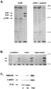
Fig. 1.
Fig. 1.
Polypeptides of 58 kDa and 53 kDa are enriched in the PSD fraction. A, Homogenate, synaptosome, and PSD fractions from rat brain were separated by SDS-PAGE, transferred to nitrocellulose, and probed with Ab98 (left) or Ab98 that had been preabsorbed with peptide (right). β-Dystroglycan is observed in the homogenate and synaptosomal fractions (β-DG). A pair of polypeptides of 58 and 53 kDa is specifically detected in the PSD fraction (p58, p53). Binding of Ab98 to all three polypeptides is eliminated when the antibody was preabsorbed (Ab98 + peptide). Mobilities of molecular weight standards are indicated. H, Homogenate;SX, synaptosomes; PSD, postsynaptic density fraction. B, Western blots of homogenate, synaptosomes, and PSD fractions from the indicated brain regions were probed with antibody Ab98. p58 and p53 are selectively enriched in the PSD fractions from all areas examined. C, Western blot of homogenate, synaptosome, and PSD fractions from cerebral cortex probed with antibodies to NMDA receptor subunit NR1, α-CaMKII, and synaptophysin.
Fig. 2.
Fig. 2.
Purification of p58 and p53 by 2D gel electrophoresis and hydrophobic interaction chromatography.A, Two-dimensional gel electrophoresis was used to separate PSD fraction proteins. Gels of equivalent samples were silver-stained (left) or blotted to nitrocellulose and probed with Ab98 (right). The positions of p58 and p53, as visualized by Western blotting, are indicated by the pair ofarrows. The migration of p58 and p53 in the first dimension indicates that these polypeptides are basic (pI, ∼9). Comparison of silver stain and Western blot shows that p58 and p53 are minor components of the PSD fraction. B, The PSD fraction proteins were solubilized in urea, loaded onto a HIC column in 1 m NaCl buffer, and then eluted in 0.1 m NaCl salt buffer. 2D gels of the HIC eluates were either silver-stained (left) or blotted to nitrocellulose and probed with Ab98 (right). p58 and p53 are readily visualized in silver-stained gels of HIC eluate (arrows), indicating that they are highly enriched by this procedure.
Fig. 3.
Fig. 3.
Structure of IRSp53. IRSp53 is predicted to contain several protein-protein interaction domains: one SH3 domain, one SH3 binding domain, and one WW binding domain (Yeh et al., 1998). Additionally, there are 25 potential serine/threonine phosphorylation sites (protein kinase A, protein kinase C, and casein kinase; data not shown) and two potential tyrosine phosphorylation sites (pY). The positions of the peptide microsequences obtained from purified p58 are noted. The positions of the epitope that is likely to be recognized by Ab98 and the peptide used to generate the polyclonal anti-IRSp58/53 antiserum are indicated. The region that corresponds to the IRSp53 DNA fragment used as a probe for Northern blots is also shown.
Fig. 4.
Fig. 4.
Relationship of IRSp58 and IRSp53 from PSDs.A, PSD fractions from rat brain were incubated under conditions promoting in vitro phosphorylation (see Materials and Methods) in either the absence (left) or presence (right) of exogenous ATP. Western blotting with Ab98 shows that IRSp58 and IRSp53 undergo similar gel shifts afterin vitro phosphorylation. B, Blots of PSD fractions from rat (left) and pig (right) were probed with antibody Ab98. Comparison of these reveals species differences in the expression of PSD fraction IRSp58 and IRSp53. Although similar amounts of IRSp58 and IRSp53 are detected in rat PSDs, only IRSp58 is detected in pig PSDs.
Fig. 5.
Fig. 5.
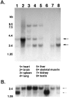
Tissue distribution of IRSp58/53 mRNAs.A, A multiple rat tissue Northern blot was probed with a radiolabeled IRSp58/53 oligonucleotide probe (248 bp; see Fig. 3) as described in Materials and Methods. Transcripts of 2.4, 3.5, and 8 kb are observed. The highest level of IRSp58/53 mRNA is detected in brain, with the 3.5 kb transcript predominating. B, The blot was rehybridized with a probe for β-actin to verify the integrity and quantity of the RNA from each tissue.
Fig. 6.
Fig. 6.
Characterization of the anti-IRSp58/53 antibody. Rabbits were immunized with a 13 amino acid peptide from the predicted amino acid sequence of IRSp58/53, and the resulting antiserum was affinity-purified. On Western blots, anti-IRSp58/53 antibody recognizes polypeptides of 58 and 53 kDa, which are also bound by Ab98. However, anti-IRSp58/53 does not recognize β-dystroglycan. All anti-IRSp58/53 immunoreactivity is abolished if the antibody is preabsorbed with peptide. H, Homogenate; SX, synaptosomes;PSD, postsynaptic density fraction.
Fig. 7.
Fig. 7.
A, Localization of IRSp58/53 in the cerebellar cortex. Sections of rat cerebellum were immunostained with the affinity-purified anti-IRSp58/53 or with anti-synapsin-I antiserum. The synapsin-I immunoreactivity reveals the distribution of synapses. IRSp58/53 immunoreactivity is observed in the synapse-rich molecular layer as well as in the granule cell layer. Anti-IRSp58/53 immunoreactivity is greatly reduced when the antibody was preabsorbed with peptide. Scale bar, 50 μm. M, Molecular layer;PC, Purkinje cell layer; GC, granule cell layer. B, Localization of IRSp58/53 at synapses. Cultured rat hippocampal neurons were immunostained with the affinity-purified anti-IRSp58/53 antiserum (left). IRSp58/53 immunoreactivity is distributed in a punctate pattern along the dendrites of the neurons. The distribution of synapses in the same dendrite was visualized by double labeling with anti-synaptophysin (right). IRSp58/53 immunoreactivity is selectively concentrated at synapses (arrows).
Fig. 8.
Fig. 8.
Insulin receptor localization in cultured neurons and brain subcellular fractions. A, Cultured rat hippocampal neurons were double-immunostained with anti-synaptophysin and anti-insulin receptor β-subunit antibody (IR-β). Insulin receptor immunoreactivity is distributed in a punctate pattern along dendrites. Note that insulin receptor β-subunit immunoreactivity is concentrated at synapses. Scale bar, 20 μm. B, High-magnification view of a single dendrite of a cultured hippocampal neuron double labeled with antibodies to the insulin receptor β-subunit and synaptophysin. IR-β immunoreactivity is concentrated at both synaptophysin-positive regions (arrowheads) as well as distributed in apparently nonsynaptic regions (arrows). Scale bar, 5 μm. C, A Western blot of rat brain subcellular fractions (homogenate, synaptosome, and PSD) and lysate from CHO.T cells was probed with an anti-insulin receptor β-subunit antibody. High levels of β-subunit are seen in CHO.T cells (a cell line engineered to overexpress insulin receptors). The β-subunit of the insulin receptor is detected in brain homogenate and is enriched in synaptosome and PSD fractions.

References

    1. Altschul SF, Gish W, Miller W, Myers EW, Lipman DJ. Basic local alignment search tool. J Mol Biol. 1990;215:403–410. - PubMed
    1. Anai M, Ono H, Funaki M, Fukushima Y, Inukai K, Ogihara T, Sakoda H, Onishi Y, Yazaki Y, Kikuchi M, Oka Y, Asano T. Different subcellular distribution and regulation of expression of insulin receptor substrate (IRS)-3 from those of IRS-1 and IRS-2. J Biol Chem. 1998;273:29686–29692. - PubMed
    1. Baltensperger K, Karoor V, Paul H, Ruoho A, Czech MP, Malbon CC. The beta-adrenergic receptor is a substrate for the insulin receptor tyrosine kinase. J Biol Chem. 1996;271:1061–1064. - PubMed
    1. Baskin DG, Schwartz MW, Sipols AJ, D’Alessio DA, Goldstein BJ, White MF. Insulin receptor substrate-1 (IRS-1) expression in rat brain. Endocrinology. 1994;134:1952–1955. - PubMed
    1. Carlin RK, Grab DJ, Cohen RS, Siekevitz P. Isolation and characterization of postsynaptic densities from various brain regions: enrichment of different types of postsynaptic densities. J Cell Biol. 1980;86:831–845. - PMC - PubMed

Publication types

MeSH terms

LinkOut - more resources