. 1999 Sep 1;19(17):7617-28.
doi: 10.1523/JNEUROSCI.19-17-07617.1999.

Mechanisms underlying spontaneous oscillation and rhythmic firing in rat subthalamic neurons

Affiliations

Mechanisms underlying spontaneous oscillation and rhythmic firing in rat subthalamic neurons

M D Bevan et al. J Neurosci. .

Abstract

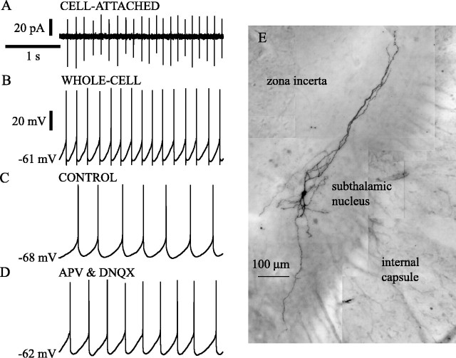
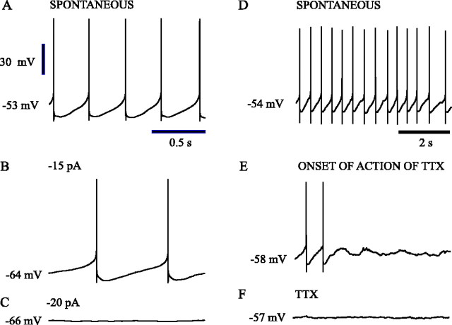
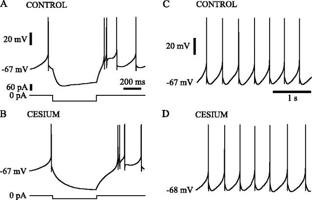
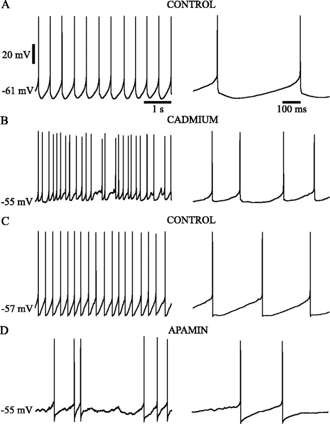
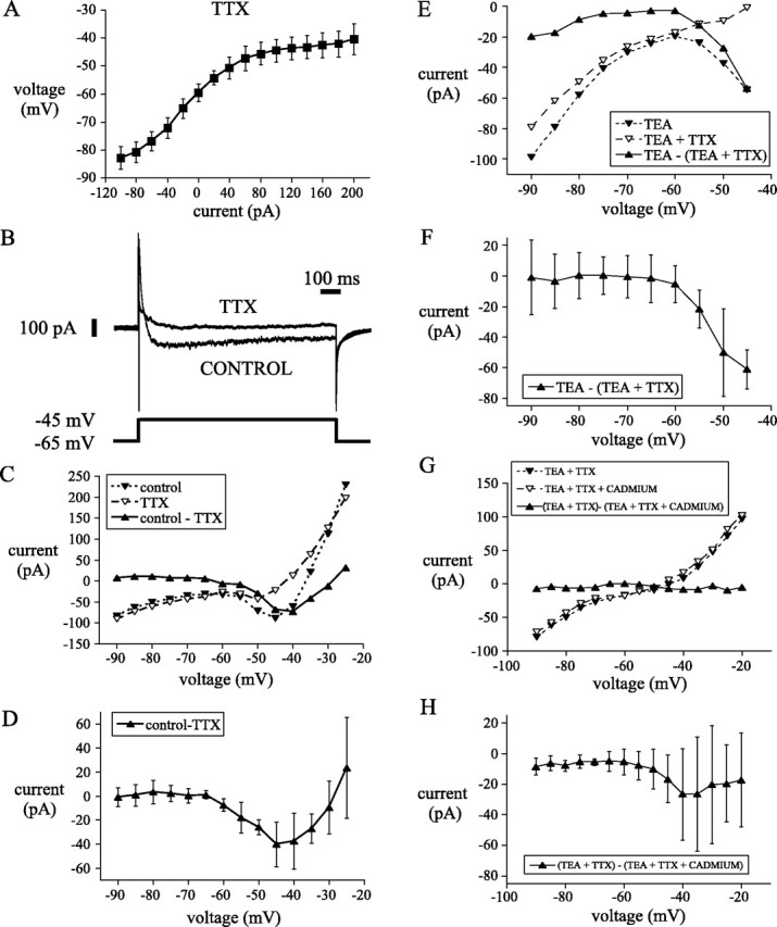
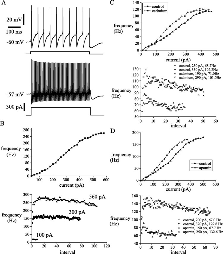
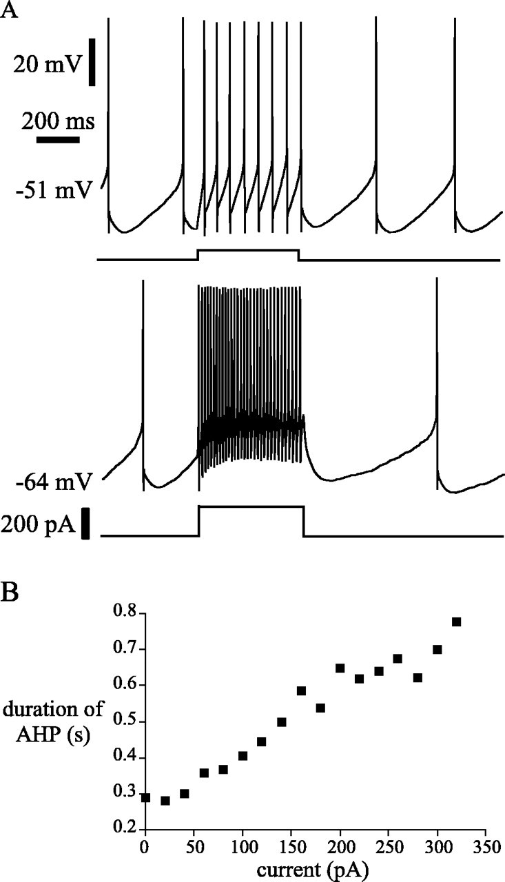
Subthalamic neurons drive basal ganglia output neurons in resting animals and relay cortical and thalamic activity to the same output neurons during movement. The first objective of this study was to determine the mechanisms underlying the spontaneous activity of subthalamic neurons in vitro and to gain insight into their resting discharge in vivo. The second objective was to determine the response of subthalamic neurons to depolarizing current injection and how intrinsic properties may shape their response to cortical and thalamic inputs during movement. Cell-attached and whole-cell recordings were made from subthalamic neurons in brain slices prepared from 3- to 4-week-old rats. The slow, rhythmic discharge of subthalamic neurons was resistant to blockade of excitatory synaptic transmission indicating that intrinsic currents underlie their spontaneous discharge. A persistent sodium current was the source of current during the depolarizing phase of the oscillation. A powerful afterhyperpolarization following each action potential was sufficient to terminate the depolarization. A long duration component of the spike afterhyperpolarization determined the period of the oscillation and was generated by an apamin-sensitive calcium-activated potassium current. Calcium entry responsible for that current was associated with action potentials. Subthalamic neurons exhibited a sigmoidal frequency-current relationship with the steeper portion starting at approximately 30-40 Hz. This property makes subthalamic neurons more sensitive to input at high firing rates associated with movement than at low rates associated with rest. We propose that the subthreshold persistent sodium current overcomes calcium activated potassium current which accumulates during high frequency firing and underlies the enhanced sensitivity to current >30 Hz.


Figures

Fig. 1.
Fig. 1.
Subthalamic neurons were spontaneously activein vitro. A, B, Example of patch-clamp recordings from a subthalamic neuron that was recorded in the cell-attached configuration (A) before establishing the whole-cell configuration (B). Note the slow (5–7 Hz), rhythmic generation of action currents(A) and action potentials (B).C, D, Intrinsic membrane properties underlie the spontaneous discharge of subthalamic neurons in vitro. A spontaneously active subthalamic neuron (C) continued to fire rhythmically when excitatory amino acid neurotransmission was blocked by the application of the selective NMDA and AMPA receptor antagonists APV and DNQX, respectively (D). E, Composite image based on multiple light micrographs of a subthalamic neuron that was visualized using histochemical procedures (see Materials and Methods) after being recorded in the whole-cell configuration. Time calibration inA applies to A–D. Current calibration inA applies to that figure. Voltage calibration inB applies to B–D. The membrane potential value printed at the left of each traces in this and all subsequent figures refers to the first point in the trace.
Fig. 2.
Fig. 2.
Sodium currents were critical to the spontaneous oscillation. Spontaneous oscillations of subthalamic neurons were abolished when action potential generation was prevented by the constant injection of negative current (A–C). The injection of negative current to a spontaneously firing subthalamic neuron (A) slowed rhythmic firing (B) before preventing it (C). When action potential generation was inhibited, underlying subthreshold oscillations were not observed (C). The application of the sodium channel blocker TTX to a spontaneously active neuron (D) abolished action potential generation (E, F), which led to a stable membrane potential at the midpoint of the voltages traversed during the subthreshold phase of the oscillation (F). Voltage calibration in Aapplies to A--F. Time calibration inA applies to A--C. Time calibration in D applies to D–F.
Fig. 3.
Fig. 3.
Hyperpolarization-activated sag current was not critical to the spontaneous oscillation. A subthalamic neuron responded to the injection of −60 pA for 500 msec with a characteristic sag in membrane potential, which was attributable to the activation of hyperpolarization-activated sag current (A). In the presence of 3 mm cesium hyperpolarization-activated sag current was blocked (B). Despite the specific block of hyperpolarization-activated sag current, the spontaneous firing of the neuron in 3 mm cesium (D) was similar to that observed in control ACSF (C). Voltage, time, and current calibrations inA also apply to B. Voltage and time calibrations in C also apply to D.
Fig. 4.
Fig. 4.
High-voltage-activated calcium currents activated potassium currents that underlie the slow single-spike afterhyperpolarization of the spontaneous oscillation. The rhythmic, spontaneous firing of subthalamic neurons (A, C) was disrupted by the application of HVA calcium current blocker (400 μm cadmium; B) or SKCa current blocker (100 nm apamin; D). High-voltage-activated and SKCa current blockade disrupted rhythmic firing by abolishing the slow single-spike afterhyperpolarization, which was activated within a few milliseconds of spike repolarization and lasted for tens or hundreds of milliseconds before the depolarizing ramp current was activated. Voltage and time calibrations inA apply to the respective parts of B andC.
Fig. 5.
Fig. 5.
A persistent sodium current was responsible for the depolarizing phase of the spontaneous oscillation.A, Steady-state I–V plot of eight subthalamic neurons recorded in current clamp in the presence of the sodium channel blocker TTX revealed no depolarizing potential in the subthreshold range of the oscillation. Inward and outward rectification were apparent at hyperpolarized and depolarized potentials, respectively. Voltage-clamp recordings were used to examine persistent currents that might underlie the depolarizing phase of the oscillation.B, A persistent TTX-sensitive current was elicited by a 1 sec step from −65 to −45 mV. C, Steady-stateI–V plot of currents elicited from the same neuron inB at the end of 1 sec steps from holding of −65 mV. Note that in control media an inward current was activated in the voltage range associated with the depolarizing phase of the spontaneous oscillation. The inward current was abolished in TTX. The TTX-sensitive sodium current (current in control media − current in TTX) shows similar voltage dependency and magnitude to the inward current observed in control media. D, The I–V plot of the persistent sodium current recorded from a population of five neurons using the same protocol exhibited a similar voltage dependence to the current in C. E-H, I–Vplots of currents elicited at the end of 1 sec steps from holding potential of −55 mV from a representative neuron (E,G) and from a sample of five neurons (F,H). Steps were made in the presence of TEA, TEA, and TTX and TEA, TTX, and cadmium. Sodium (TTX-sensitive) current was obtained by subtraction of currents in TEA and TTX from currents in TEA. High-voltage-activated calcium (cadmium-sensitive) current was obtained by subtraction of currents in TEA and TTX from TEA, TTX, and cadmium. Note that the persistent sodium current is activated in the subthreshold range of the oscillation (E, F), whereas persistent calcium currents are smaller and less reliable in the subthreshold range (G-H).
Fig. 6.
Fig. 6.
Subthalamic neurons fired rhythmically with minimal spike frequency adaptation in response to current injection and exhibited a sigmoidal f–I relationship.A, Example of driven rhythmic firing by a subthalamic neuron. B, The same neuron displayed a sigmoidalf–I relationship. Note the transition to secondary range firing occurred at ∼35 Hz. Subthalamic neurons exhibited a rapid speed-up in instantaneous firing frequency in the first few intervals of a driven spike train in the secondary and tertiary ranges. From the point of maximal firing frequency a small spike frequency adaptation developed slowly. C, D, Calcium-activated potassium current limited excitability of subthalamic neurons during high-frequency firing. Suppression of calcium-activated potassium currents with cadmium (C) or apamin (D) shifted the f–I relationship to the left and disrupted low-frequency firing associated with the primary range. Speed-up and spike frequency adaptation within driven trains of spikes were present when calcium and SKCacurrents were blocked.
Fig. 7.
Fig. 7.
Saturation of afterhyperpolarization did not account for the sigmoidal f–I relationship of subthalamic neurons because afterhyperpolarization accumulated during high-frequency firing. A, B, Representative example of a spontaneously firing subthalamic neuron, which was injected with an increasing magnitude of current. Note that the time to the resumption of spontaneous firing, a measure of afterhyperpolarization, increased as both elicited firing frequency and driving current increased.

References

    1. Alzheimer C, Schwindt P, Crill W. Modal gating of Na+ channels as a mechanism of persistent Na+ current in pyramidal neurons from rat and cat sensorimotor cortex. J Neurosci. 1993;13:660–673. - PMC - PubMed
    1. Benazzouz A, Gross C, Feger J, Boraud T, Bioulac B. Reversal of rigidity and improvement in motor performance by subthalamic high-frequency stimulation in MPTP-treated monkeys. Eur J Neurosci. 1993;5:382–389. - PubMed
    1. Benazzouz A, Boraud T, Feger J, Burbaud P, Bioulac B, Gross C. Alleviation of experimental hemiparkinsonism by high-frequency stimulation of the subthalamic nucleus in primates: a comparison with L-Dopa treatment. Mov Disord. 1996;11:627–632. - PubMed
    1. Bennett BD, Wilson CJ. Synaptic regulation of action potential timing in neostriatal cholinergic interneurons. J Neurosci. 1998;18:8539–8549. - PMC - PubMed
    1. Beurrier C, Congar P, Bioulac B, Hammond C. Subthalamic neurons switch from single-spike activity to burst-firing mode. J Neurosci. 1999;19:599–609. - PMC - PubMed

Publication types

MeSH terms

LinkOut - more resources