

Microbial decontamination of human donor eyes with povidone-iodine: penetration, toxicity, and effectiveness
- PMID: 10460768
- PMCID: PMC1723175
- DOI: 10.1136/bjo.83.9.1019
Microbial decontamination of human donor eyes with povidone-iodine: penetration, toxicity, and effectiveness
Abstract
Background/aims: Povidone-iodine (PVP -I) is applied for microbial decontamination of human eyes donated for transplantation. Concentrations and immersion times vary greatly. The effectiveness and toxicity of PVP-I were assessed for different decontamination protocols.
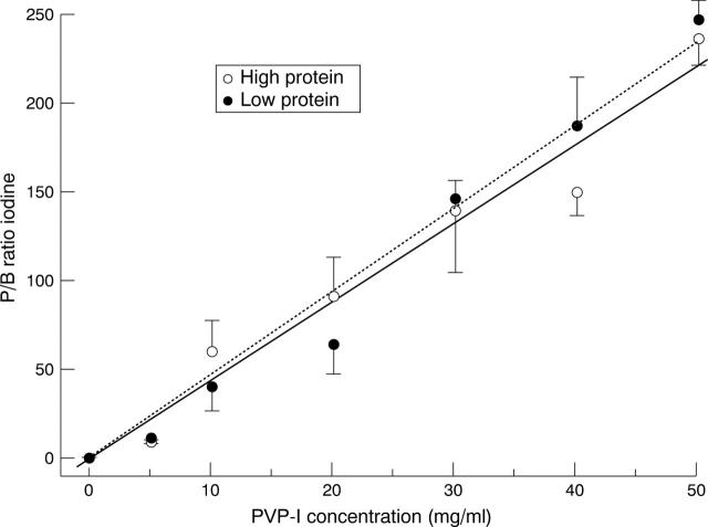
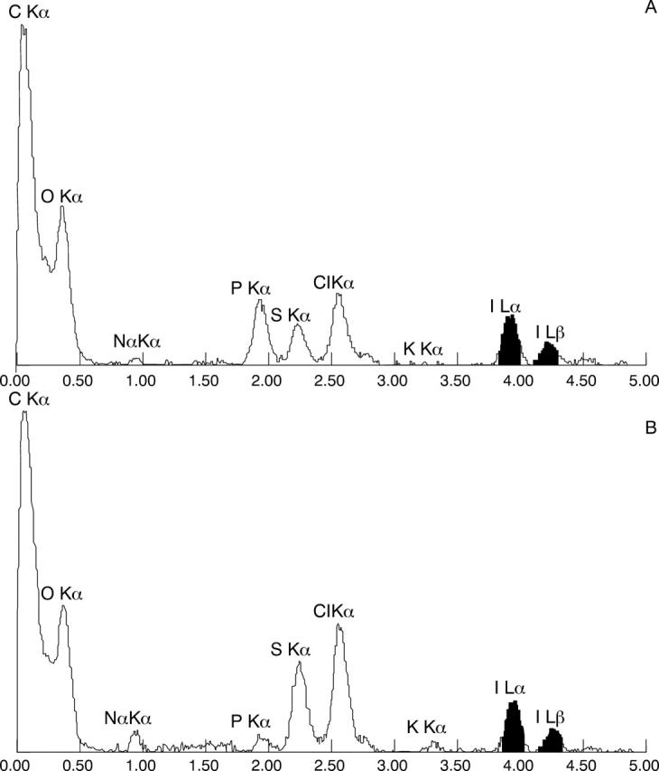
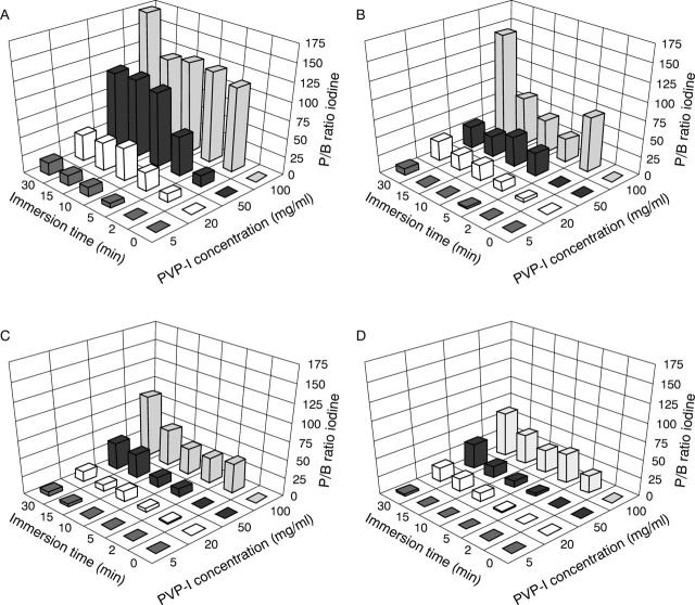
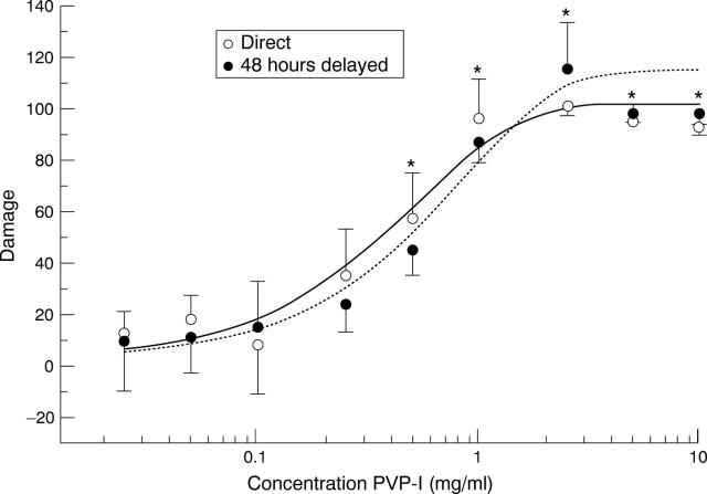
Methods: Human donor eyes and corneas were immersed in different concentrations (5-100 mg/ml) of PVP-I for different times (2-30 minutes). The penetration of iodine into the corneal tissue was assessed by x ray microanalysis. Microbial contamination was determined by taking cultures of the limbal areas and storage solutions and by incubation of the corneoscleral buttons in antibiotic-free culture medium. Cytotoxicity of PVP-I for corneal fibroblasts in culture was assessed using the MTT assay.
Results: Depending on concentration and immersion time iodine was found to penetrate into the epithelium, Bowman's layer, and stroma in amounts equivalent to 2-40 mg/ml PVP-I. The MTT assay demonstrated that 2.5 mg/ml PVP-I caused total damage to fibroblasts in vitro. Rinsing eyes with tap water and subsequent immersion in PVP-I reduced the rate of contamination from 82 out of 106 to 69 out of 106 and 37 out of 106, respectively. Antibiotics in the storage medium further reduced contamination from about 40% to 3%. Microbial contamination was not reduced by increasing the concentration and immersion times beyond 5 mg/ml PVP-I for 2 minutes.
Conclusion: Immersion of human donor eyes in 5 mg/ml PVP-I solution for 2 minutes significantly reduces microbial contamination of donor corneas without relevant penetration of iodine into the corneal layers. Higher PVP-I concentrations and longer immersion times do not further reduce contamination, whereas the amount of iodine penetrating the corneal layers is elevated above the level cytotoxic for corneal fibroblasts. In view of this, concentrations above 5 mg/ml of PVP-I and immersion periods over 2 minutes are not recommended for reduction of the contamination rate of donor eyes.
Figures

References
MeSH terms
Substances
LinkOut - more resources
Full Text Sources
Other Literature Sources
Miscellaneous