. 1999 Sep;181(17):5185-92.
doi: 10.1128/JB.181.17.5185-5192.1999.

Amino acid-DNA contacts by RhaS: an AraC family transcription activator

Affiliations

Amino acid-DNA contacts by RhaS: an AraC family transcription activator

P M Bhende et al. J Bacteriol. 1999 Sep.

Abstract

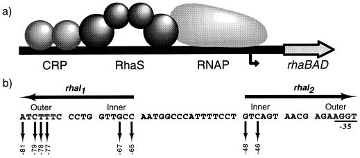
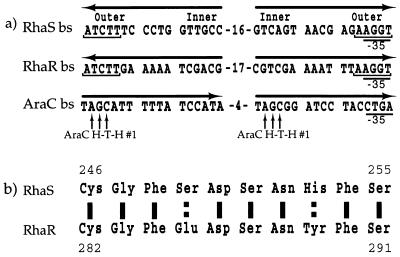
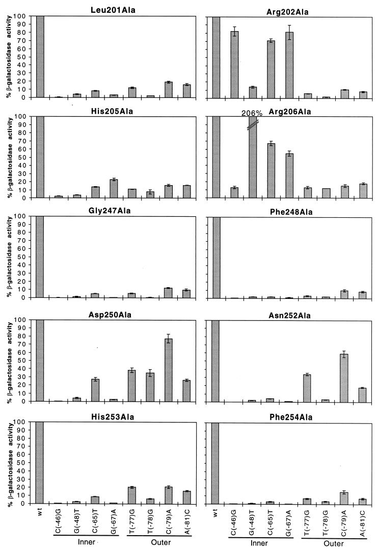
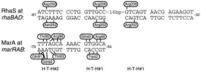
RhaS, an AraC family protein, activates rhaBAD transcription by binding to rhaI, a site consisting of two 17-bp inverted repeat half-sites. In this work, amino acids in RhaS that make base-specific contacts with rhaI were identified. Sequence similarity with AraC suggested that the first contacting motif of RhaS was a helix-turn-helix. Assays of rhaB-lacZ activation by alanine mutants within this potential motif indicated that residues 201, 202, 205, and 206 might contact rhaI. The second motif was identified based on the hypothesis that a region of especially high amino acid similarity between RhaS and RhaR (another AraC family member) might contact the nearly identical DNA sequences in one major groove of their half-sites. We first made targeted, random mutations and then made alanine substitutions within this region of RhaS. Our analysis identified residues 247, 248, 250, 252, 253, and 254 as potentially important for DNA binding. A genetic loss-of-contact approach was used to identify whether any of the RhaS amino acids in the first or second contacting motif make base-specific DNA contacts. In motif 1, we found that Arg202 and Arg206 both make specific contacts with bp -65 and -67 in rhaI1, and that Arg202 contacts -46 and Arg206 contacts -48 in rhaI2. In motif 2, we found that Asp250 and Asn252 both contact the bp -79 in rhaI1. Alignment with the recently crystallized MarA protein suggest that both RhaS motifs are likely helix-turn-helix DNA-binding motifs.


Figures

FIG. 1
FIG. 1
(a) Model for transcription activation at rhaBAD, showing the two activator proteins, cyclic AMP receptor protein (CRP) and RhaS, and RNA polymerase (RNAP) shown. The bent arrow represents the rhaBAD transcription start site. (b) DNA sequence of the RhaS binding site (rhaI). The two half-sites, rhaI1 and rhaI2, are inverted repeats, as indicated by the arrows above the sequence. Sequences within the half-sites are divided into two major grooves separated by a minor groove, with the outer and inner major grooves indicated. Down arrows indicate bases previously identified as important for interaction with RhaS (9); their positions relative to the rhaBAD transcription start site are indicated below. Bases overlapping the −35 region of the promoter were not analyzed in this previous study since their effects on RhaS binding and RNA polymerase binding could not easily be distinguished by the analysis used. Notice that all of the bases important in rhaI1 are identical in rhaI2.
FIG. 2
FIG. 2
(a) Comparison of the DNA-binding sites (bs) recognized by RhaS, RhaR, and AraC. Horizontal arrows indicate the half-sites and their relative orientations. Notice that the downstream half-sites are all in the same relative orientation whereas the upstream half-sites are not. Bracketed bases are conserved in the RhaS and RhaR DNA-binding sites. The outer and inner major grooves of the RhaS binding site are indicated. The −35 regions of the promoters are shown. Vertical arrows indicate amino acid-base pair contacts between AraC H-T-H 1 and its DNA-binding site. (b) Sequence of amino acids 246 to 255 of RhaS, showing high similarity with the aligned region of RhaR (amino acids 282 to 291). Solid lines indicate identical amino acids; broken lines represent similar amino acids.
FIG. 3
FIG. 3
Alanine substitutions in RhaS H-T-H 1 and 2 analyzed at mutant rhaB-lacZ fusions. The x axis (labeled at the bottom) represents either the wild-type rhaBAD promoter or the position of point mutations in rhaI found to be important for RhaS binding (Fig. 1). Locations of the point mutations in either the inner or outer major grooves of rhaI are indicated. The y axis (labeled on the left) represents the percent β-galactosidase specific activity for each RhaS alanine mutant at mutant rhaB-lacZ compared with the same mutant protein at the wild-type rhaB-lacZ fusion. The first bar in each graph represents the RhaS alanine mutant protein assayed at the wild-type rhaB-lacZ promoter and is set to 100%. Error bars are shown.
FIG. 4
FIG. 4
Summary of specific amino acid-base pair contacts by RhaS and comparison with MarA DNA contacts. DNA half-sites are divided into two major grooves and an intervening minor groove. Vertical and angled black lines represent amino acid-base pair contacts. The broken bar indicates a less conclusive DNA contact by RhaS. The side of the DNA sequence on which each amino acid is represented is arbitrary and is not meant to represent the base within the pair that is contacted. For MarA, the base(s) within each pair that is contacted is known (30); however, this information is not represented for simplicity and because the corresponding information is not known for RhaS. Relative positions of H-T-H 1 and 2 are shown below the two DNA sequences.

References

    1. Backman K, Chen Y-M, Magasanik B. Physical and genetic characterization of the gln A-glnG region of the Escherichia coli chromosome. Proc Natl Acad Sci USA. 1981;78:3743–3747. - PMC - PubMed
    1. Bourgerie S J, Michan C M, Thomas M S, Busby S J W, Hyde E I. DNA binding and DNA bending by the MelR transcription activator protein from Escherichia coli. Nucleic Acids Res. 1997;25:1685–1693. - PMC - PubMed
    1. Brunelle A, Schleif R. Determining residue-base interactions between AraC protein and araI DNA. J Mol Biol. 1989;209:607–622. - PubMed
    1. Brunelle A, Schleif R. Missing contact probing of DNA-protein interactions. Proc Natl Acad Sci USA. 1987;84:6673–6676. - PMC - PubMed
    1. Chiang L W, Kovari I, Howe M M. Mutagenic oligonucleotide-directed PCR amplification (Mod-PCR): an efficient method for generating random base substitution mutations in a DNA sequence element. PCR Methods Appl. 1993;2:210–217. - PubMed

Publication types

LinkOut - more resources